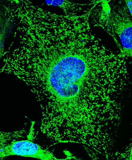
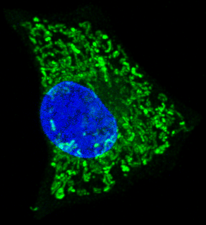
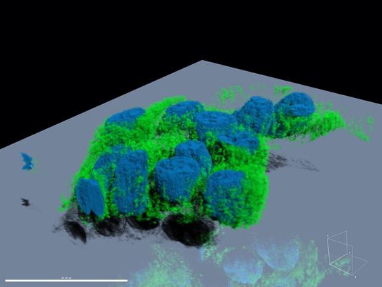
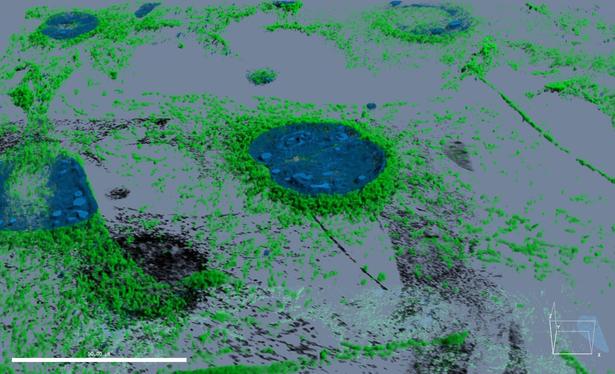
Had to revisit our paper from 2018 for a future project…boy I love the immunofluorescence microscopy we did on the human placenta.
#FluorescenceFriday #Science #Placenta #ReproSci
https://bmcdevbiol.biomedcentral.com/articles/10.1186/s12861-018-0178-0
#FluorescenceFriday
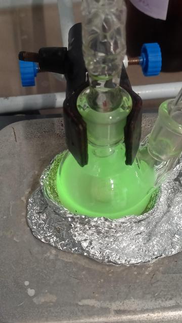
terbium something (idk?) #fluorescencefriday
Linda semana, sumergido en el mundo microscópico…
🔬🦠
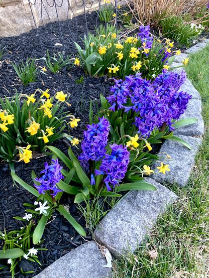
Front yard😊
Not really fluorescence, but like fluorescence 😉
For #fluorescencefriday I present: stoma falling into the abyss, on a dying flower. The other tech gave me beautiful purple flowers for my birthday and I just had to take a look. In microscopy I’m always interested how things tear/break/rip and otherwise fall apart... #microscopy #sciart
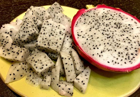
Some nice colors of Dragon fruit 😊😋
This reminds me that I should plan some fluorescence microscopy experiments 🤔
#DragonFruit #Fruits #Photography #Fluorescence #Science #Experiment
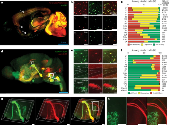
New publication from Kwanghun Chung lab at MIT improves on their SWITCH approach and eFLASH stochastic electrotransport to improve antibody labeling by gradually shifting the microenvironment. Looks interesting, I wonder how well it can be adapted to other labeling protocols.
Uniform volumetric single-cell processing for organ-scale molecular phenotyping
Yun et al., Nature Biotechnology 2025
https://doi.org/10.1038/s41587-024-02533-4
#neuroscience #tissueclearing #lightsheet #microscopy #fluorescenceFriday
I missed #FluorescenceFriday but here's an image for one from the manuscripts on the nearly-finished pile. We've found a genetic marker for the deep CA1 pyramidal cells. Magenta is calbindin (IHC), cyan is the other cell type (AAV-TdTom). Less than 5% overlap. Coming to BioRxiv by Easter, hopefully! #Neuroscience
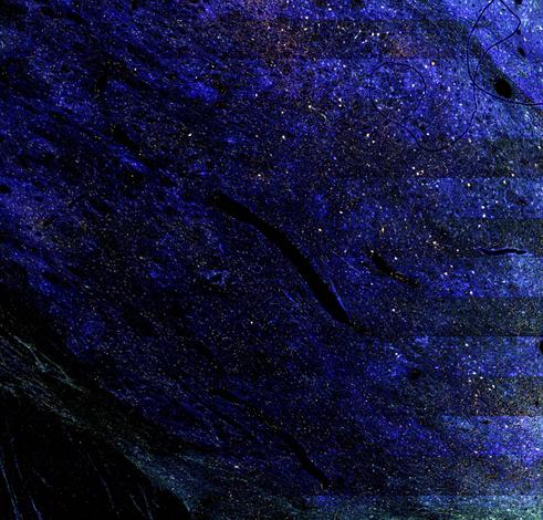
Night sky in the thalamus. Or is it cities from high above? Fluorescent microscopy is my fave so I’m super happy to be on the confocal this week. #fluorescencefriday #neurons #sciart
You can check it out here: https://doi.org/10.1016/j.jid.2024.10.595
#fluorescencefriday every day...
We are especially grateful to CMN patient advocacy groups, MELCAYA, AFM-Téléthon, and the Institut Marseille Maladies Rares (MarMaRa), for their support.
Bon bout d'an à tous ! (Happy nearly-New Year to all!)
-fin-
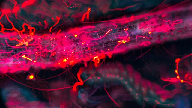
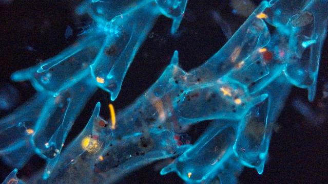
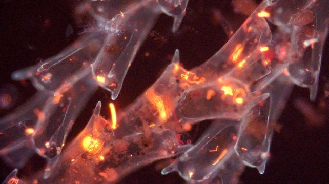
My contribution to #FluorescenceFriday is this mysterious hot pink plant debris with algae on it, so many little shapes and details! Along with the trails of flagellates (orange) and cladocerans (blue-green). #SciArt #chronophotography #microscopy #daphnia
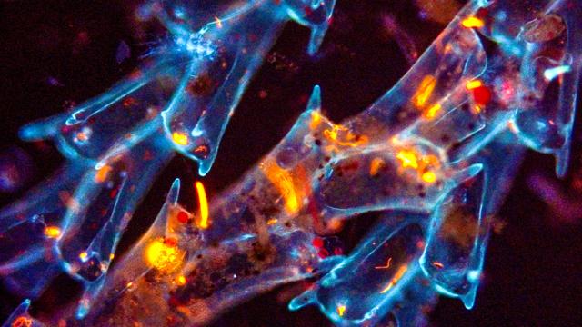
Is #FluorescenceFriday still a thing? I recently made my very first youtube video- in it I compare my triple bandpass fluo filter to a UV longpass filter imaging autofluorescence of plankton: youtu.be/n2rCXnJ50Y8?... Love having a fluo setup! Below, decomposing algae in both filters. #SciArt
@MFM_BIC@synapse.cafe @MFM_BIC
ICYMI you can follow tags in here:
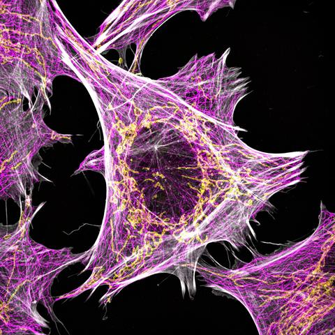
On #FluorescenceFriday, we’re highlighting this beautiful SIM image from @dgaboriau These human fibroblasts are labelled to visualise mitochondria (Tom20) in yellow, alpha-tubulin in pink and actin in grey.
https://focalplane.biologists.com/2024/10/11/featured-image-with-david-gaboriau/
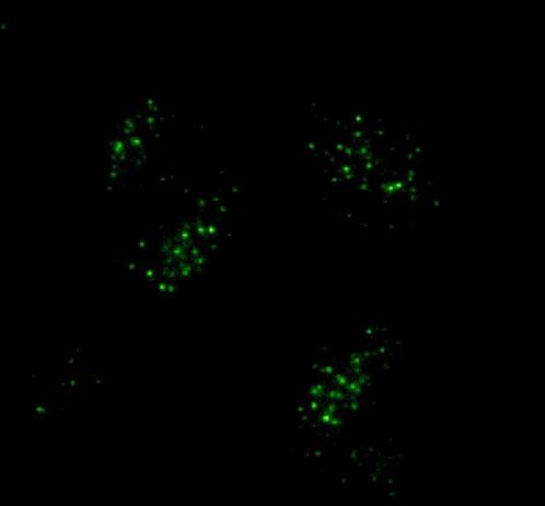
Some nice telomere signals, I think experiments are working
#Telomere #Fluorescence #CellBiology #MolecularBiology #GraduateSchool
for #FluorescenceFriday, here's a video of two-photon imaging 🔬 from mouse visual cortical neurons at 0.5x speed in response to many natural images #neuroscience (paper here: https://www.biorxiv.org/content/10.1101/2024.06.30.601394v1)
Happy #fluorescenceFriday, let’s see those gorgeous images!
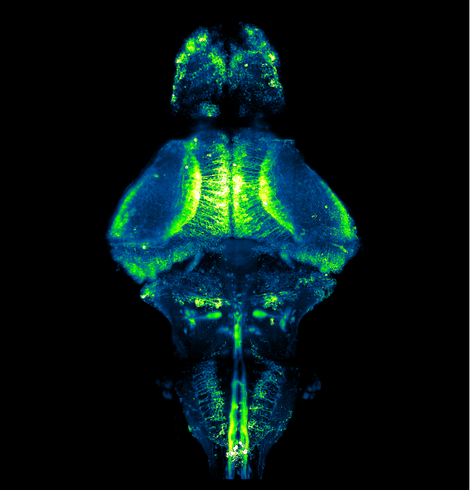
It’s #FluorescenceFriday & we’re highlighting Shivangi Verma’s (@shivangiv10) image of a #zebrafish larval brain. The larva expressing GCaMP6f was embedded in agarose & imaged using a Zeiss Lightsheet microscope. Find out more about Shivangi's research:
https://focalplane.biologists.com/2024/08/30/featured-image-with-shivangi-verma/
Happy #FluorescenceFriday
Some actin action always - amazes me (and gets me safely into the weekend) 😜
Timelapse imaging on Zeiss880 of HEK cells expressing Lifeact-mEGFP
LUT is Cyan Hot (quite like that one)
#FluorescenceFriday
(is this a thing on #Mastodon - if not let's make it one!)
Nuclei labelled with EGFP-H2B.
MIP of a small z-stack at two frames per minute.
Loads of action, movement and cell divisions, in the developing zebra fish hindbrain 🐟🧠
