#lightmicroscopy
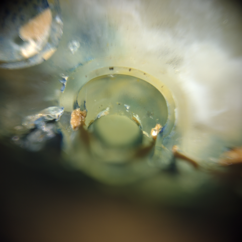
Image width is 3 millimeters 📏 and obtained by white light reflection.
#science #mineraloid #opal #lightmicroscope #lightmicroscopy #microscope #microscopy #microscopyart #lens #doityourself #doityourselfexperiment #buildityourself
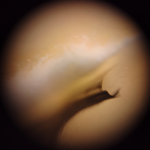
Image width is 3 millimetres 📏 and obtained by white light reflection.
#science #mineral #mineralogy #agate #lightmicroscope #lightmicroscopy #microscope #microscopy #microscopyart #lens #doityourself #doityourselfexperiment #buildityourself
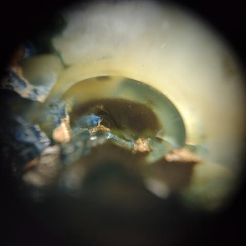
Image width is 3 millimeters 📏 and obtained by white light reflection.
#science #mineraloid #opal #lightmicroscope #lightmicroscopy #microscope #microscopy #microscopyart #lens #doityourself #doityourselfexperiment #buildityourself
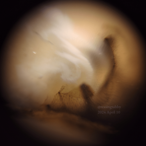
Image width is 3 millimetres 📏 and obtained by white light reflection.
#science #mineral #mineralogy #agate #lightmicroscope #lightmicroscopy #microscope #microscopy #microscopyart #lens #doityourself #doityourselfexperiment #buildityourself #wavy #creamy
The NeuronBridge website, for matching #ElectronMicroscopy and #LightMicroscopy data for #Drosophila #Neuroscience research, now includes EM data from FlyWire. Getting it working was a great effort by @neomorphic, Cristian Goina, Hideo Otsuna, Robert Svirskas, and @konrad_rokicki. Here is a screen capture of looking up a neuron on FlyWire Codex, finding its match in NeuronBridge, then viewing the match in 3D with volume rendering in the browser.
https://www.nature.com/articles/d41586-024-03276-7
"Researchers are queuing up to try a powerful microscopy technique that can simultaneously sequence an individual cell’s DNA and pinpoint the location of its proteins with high resolution — all without having to crack the cell open and extract its contents"
😮🔬🧬
Image width is 3 millimeters 📏 and obtained by white light reflection
#science #agate #mineral #mineralogy #lightmicroscope #lightmicroscopy #microscope #microscopy #microscopyart #lens #doityourself #doityourselfexperiment #buildityourself
Image real width is 3 millimeters 📏.
#agate #mineral #mineralogy #lightmicroscope #lightmicroscopy #microscope #microscopy #lens #science #doityourself #buildityourself #wavy #creamy
Are you doing cell biology or biomedical research? Light microscopy-based imaging? If a facility for (multiplexed) imaging would pop up in your department, what would you wish for? If you (would) run one, what is crucial?
I’ve applied for a (multiplexed) imaging facility manager job and would like to collect a few use scenarios (think tools, training, data analysis) to prepare for the interview.
#CellBiology #Microscopy #Imaging #Biology #LightMicroscopy #Science #academicchatter #academia
⚡️ Deadline extension ⚡️ You can still apply for #EMBLWidefieldConfocal 🙌
Apply by 9 February and learn how to obtain the best results when using light microscopy!
➡️ https://s.embl.org/mic24-01
📍 EMBL Heidelberg, Germany
📅 22 – 26 April
👥 This course is ideal for PhD students and faculty searching for instruction on light microscopy and basic image processing, with a basic level of microscopy.
#lightmicroscopy #lifesciencetraining #microscopytraining #microscopycourse #imageprocessing
Want to know how to attain the best results when using light microscopy? 🔬💡 Then join our hands-on #EMBLWidefieldConfocal course to learn the basics and get to know the image analysis tools needed!
Apply by 29 January 👉 https://www.embl.org/about/info/course-and-conference-office/events/mic24-01/
🗺️ EMBL Heidelberg
📅 22 – 26 April
🤝 This EMBL course is co-organised with Evident Scientific, so participants will also get the chance to connect with industry experts during the course.
1/6
Hello to the #ISMRM23 greetings from Germany! Our group is presenting today and tomorrow
#hMRI #microstructure #DWI #MPMs #AxonRadii #MRgratio #DeepLearning #Lightmicroscopy #epilepsy
Hi fellow humans, I am Laura (zie/zir if possible, otherwise she/her), a senior staff scientist at #LSFOctopus advanced #LightMicroscopy and #CLEM facility at #HarwellCampus in the #UK, right next to #DiamondLightSource.
My science interest center around #EGFRSignalling and #RTKSignalling and deregulation in #CancerStudies in general. My tools of the trade are #singleparticletracking #singlemoleculelocalisation #CLEM #FIBSEM #VolumeEM and #CryoMicroscopy
HMU for science chat


