GlycoShape

GlycoShape is an OA database of glycans 3D structural data and information that can be downloaded and/or used with Re-Glyco to rebuild glycoproteins from the RCSB PDB or EMBL-EBI AlphaFold repositories or own. GlycoShape can also predict N-glycosylation occupancy with GlcNAc Scanning. Developed and curated by Elisa Fadda research group @Elisa

GlycoShape boosted:
NCFGncfg
2025-01-15

Our latest blog on interesting facts about glycans we dig deeper into the UDP-glucuronic acid and glucuronyltransferases in the endoplasmic reticulum - which can be critical in drug metabolism.

Read at: research.bidmc.org/ncfg/blog/f

2024-11-08

*Note: pun intended 😜

2024-11-08

A spoonful of sugar #18 🍬

Plant receptor kinases translate and pass on chemical messages as part of many key biological processes. A new fabulous cryo-EM structure of the active complex between the kinase MIK2, the immunomodulatory peptide SCOOP and the co-receptor BAK1 shows how its binding hinges on N-glycosylation (doi.org/10.1038/s41477-024-018)

You can rebuilt the fully glycosylated structure of the complex with glycoshape.org with some fancy-plant N-glycans :awesome:

#glycotime

Structure of the complex between MIK2 (green), the immunoregulatory peptide SCOOP (yellow) and the he extracellular Leu-Rich-Repeat of the co-receptor BAK1 (BAK1LRR) in white. the part of the N-glycans resolved are shown with sticks (C atoms in cyan, O in red and N in dark blue) PDBid 8AYY (Cryo-EM, 3.34 Å resolution)
2024-11-06

@MissGayle We are based in the UK, luckily! But we are always making plans :P

2024-11-06

On this page we are focusing on #science, yet we are not immune to the political and social environment around all of us

We are a small research group interested in all about sugar biology 🍬 structural biology 🌱 (see above) and computational biophysics πŸ’»

We operate *exclusively* here on #mastodon #fediverse, where the scientific discourse needs to be better supported

We think we are doing our bit, please support (πŸ‘+ReTooT) #science posts to amplify scientific content on free servers!

2024-11-06

A spoonful of sugar #17 🍬

Everyone needs a bit of sugar today, here is a treat

The oligosaccharyltransferase, or OST (EC 2.4.1.119), is a key enzyme that functionalizes nascent proteins with sugars called N-glycans

The 3D structure of this magnificent molecular machine has been solved by cryo-EM by Ana Ramirez et al. (PDB 8AGC) doi.org/10.1038/s41467-022-350

Be like OST and plant one of the 195 N-glycans 🌱 online on your protein 3D structure with glycoshape.org :awesome:

#glycotime

Reduced model of the OST (STT3 and part of the OST3 domains only) shown in grey, substrate peptides shown in purple and lipid bilayer (ER membrane) model shown in green
2024-10-25

A spoonful of sugar #16 🍬

How does glycosylation affect not only structure and function of proteins, but also binding specificity?

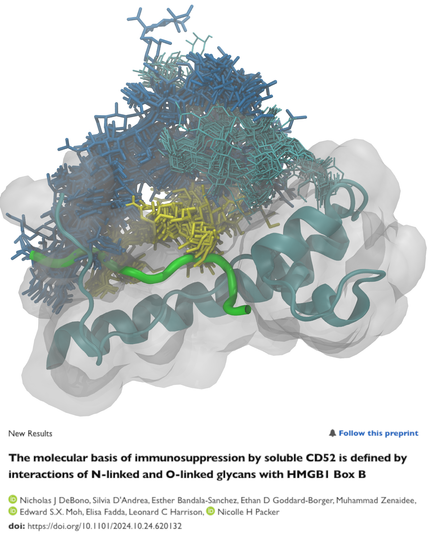
In a new paper fresh off the #bioRxiv press doi.org/10.1101/2024.10.24.620 in collaboration with Nicki Packer, Len Harrison and Ethan Goddard-Borger, we explain how glycosylation of CD52 (reconstructed with glycoshape.org) determines:
1) the peptide's conformation,
2) binding to the HMGB1 Box B and only then
3) to SIGLEC-10 :awesome:

#glycotime

Graphical representation of the 3D model of the complex between CD52 (green) O-glycosylated (yellow) and N-glycosylated (blue) to HMGB1 BoxB (cyan cartoons and white transparent surface) also N-glycosylated (thinner cyan sticks). Title of the paper below with authors list: 

New Results A Follow this preprint The molecular basis of immunosuppression by soluble CD52 is defined by interactions of N-linked and O-linked glycans with HMGB1 Box B by Nicholas  DeBono, Silvia D'Andrea, Esther Bandala-Sanchez, Ethan D Goddard-Borger, Muhammad Zenaidee, Edward S.X. Mo, Elisa Fadda, Leonard C Harrison, Nicolle H Packer doi: https://doi.org/10.1101/2024.10.24.620132
2024-10-14

6/6 Last but not least GlycoShape was and continues to be developed for and with the #glycotime community :blobcheerbounce: BIG MASSIVE THANK YOU πŸ‘ to all the beta testers around the world. Happy glycosylating! :awesome:

graphical representation of the glycoshape research team withe everyone represented by their favourite monosaccharide symbol from the SNFG. Concept by O. Singh and rendering by DALL-E
2024-10-14

5/6 GlycoShape was developed with the generous support of ORACLE for Research and funded by the Science Foundation of Ireland (SFI) Additional resources provided by ICHEC and amazing ongoing support from IRIDIS HPC at the University of Southampton

bluish image of a computer card from creative commons
2024-10-14

4/6 GlycoShape (glycoshape.org) is an OA database and toolbox that allows you to restore the 3D structure of glycoproteins with a fast and accurate algorithm named Re-Glyco.

The database counts > 550 glycans to date, largely from the human glycome with examples from all species. For an overview check out the figure below ⬇️

graphical representation of the structure and capabilities running on GlycoShape and ReGlyco
2024-10-14

3/6 Analogously, glycoproteins carry beautifully complex sugars, with precise structures and functions, yet invisible due to their fast dynamics and heterogeneous nature

electron density map from PDB 5GQR with partially resolved sugars
2024-10-14

2/6 What you see through a camera lens depends on the shutter speed.

Yet we all know that hummingbirds do have wings, and that these are precisely shaped to allow the fast and complex dynamics crucial to their ability to hover and feed on nectar

picture of a hummingbird in flight, the wings are moving too fast relative to the camera shutter speed and thus cannot be seen. Original photo by Christina Doherty on threads.net
2024-10-14

Some exciting #glycotime to start the week!

GlycoShape is fresh off the press today on Nature Methods nature.com/articles/s41592-024 :awesome:

Check out the short thread below 1/6

#Science #ProteinStructure #glycobiology

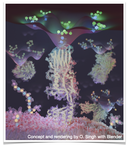
Graphic representation of the process implemented into GlycoShape to restore the sugars (glycans) on the 3D structures of key glycoproteins in human health and disease, as well as synthetic systems uploaded by users, top left. All glycans are shown according to the Symbol Nomenclature for Glycans (SNFG). Graphic concept and rendering by O. Singh with Blender.
2024-10-09

The Nobel Prize in Chemistry 2024 goes to David Baker (1/2) and Demis Hassabis and John Jumper (1/2) for protein structure design and prediction from sequence with AI

nobelprize.org/prizes/chemistr

Congratulations to all very worthy winners from all of us! πŸ‘ πŸ₯³

As a reminder, just add sugars with glycoshape.org :awesome:

#glycotime #ChemNobel #Chemistry #Science

Official portraits of the Nobel Prize Laureates by Niklas Elmehed, Left David Baker, centre Demis Hassabis and Left John M Jumper
2024-10-08

A spoonful of sugar #15 🍬

Arabinose is an aldopentose (five-membered ring sugar terminating with a CHO group) commonly found in plants as part of chains O-linked to hydroxy-Pro (Hyp) residues in the protein

As an example, look below at the magnificent structure of an algal mastigoneme resolved by cryo-EM (Dai et al. doi: 10.1016/j.cell.2024.03.005)

For all your arabinose (and Galf) needs, go to glycoshape.org , we got you covered! :awesome:

#glycotime

cryo-EM structure of the algal mestigoneme filament MST3 from the paper by Dai et al DOI 10.1016/j.cell.2024.03.005. This is a screenshot from Figure 3, showing the density with the structures of the Ara-chains that could be fitted in.
2024-08-02

🚨 Part 2/2

In view of the next update we are extending the database to contain examples of glycan structures from bacteria, plants and important model organisms

Look ⬇️ at these super fancy fucosylated N-glycan forms from C. elegans from the brilliant work by K. Paschinger, @iainwilson and team (see DOIs doi.org/10.1016/j.mcpro.2023.1 and doi.org/10.1016/j.jbc.2023.103) brewing now on our HPCs :awesome:

Four starting structure (green run1, cyan run2, purple run3 and blue run 4) of a fancy bisected N-glycan found in C elegans (see main post for refs). These structures were built with the carbohydrate builder available OA at glycam.org 

3D structures on the left and SNFG 2D representation on the right with linkage info (obtained from http://www.virtualglycome.org/DrawGlycan/)
2024-08-02

A spoonful of sugar #14 🍬

🚨 Part 1/2

We have updated the GlycoShape database that now counts 453 3D glycan structures!

Check it out at glycoshape.org :awesome:

One of the main changes is that now all glycans are listed by their GlyTouCan IDs, a unique identifier that allows you to search all data related to your favourite glycan across the GlyCosmos (DOI 10.1093/glycob/cwx066) 😎

A few structures still do not have GlyTouCan IDs, we are currently registering them

#glycotime

GlyTouCan Logo. GlyTouCan is named by a combination of β€œGlycan” and β€œTou (η³–)”, which means β€œsugar” in Japanese. The word β€œCan (Jpn. ηΌΆ)” of GlyTouCan signifies that it is a container where glycans are accumulated. Therefore, a can is used in the logo to indicate that it is a repository with accumulated glycan data. In addition, a toucan is used as a mascot character since the word β€œTouCan” is included in GlyTouCan. The GlyTouCan toucan sits, appropriately, on the branch of a Lewis type glycan structure. This figure is sourced from the original paper Tiemayer et al, Glycobiology (2017) doi: 10.1093/glycob/cwx066
2024-07-26

A spoonful of sugar #13 🍬

Our team continuously updates the glycan structure database on glycoshape.org with new glycans, through a process we call glycan of the week (GOTW) :awesome:

Every week each one of us has (at least) one glycan of their choice to run and analyse through a strict MD protocol for high-quality standards. The results are progressively added to the database.

If you would like your fav glycan in the database, let us know via comment or by email 😎

#glycotime

example of a GOTW with glycan represented in 2D (top) and 3D (bottom) through SNFG colour scheme and symbols
GlycoShape boosted:
NCFGncfg
2024-06-13

This month's interesting fact about glycoscience is about made by bacteria and the importance of in antibiotics.

Read the full post at: research.bidmc.org/ncfg/blog/f

For more interesting facts check: research.bidmc.org/ncfg/blog

2024-06-13

A spoonful of sugar #12 🍬

Glycans are an integral part of a glycoproteins structure and function, as shown in our most recent paper doi.org/10.7554/eLife.95708.3 showing how N-glycosylation allosterically regulates the ability of the SARS-CoV-2 S RBD to bind co-receptors

This ability is regulated by mutations, which can in turn allow viral evolution to alter the shield and the virus' immune profile

All glycans added with glycoshape.org :awesome:
#glycotime

In yellow the 3D structure of the SARS-CoV-2 spike (S) receptor binding domain (RBD) in the omicron BA.1 variant, rendered with an opaque surface. The surface becomes clear to highlight the structural function  of the N-glycan at N343, highly conserved glycosylation site in the SARS-CoV-2 phylogeny (see Harbison et al. https://doi.org/10.1039/D1SC04832E). The glycan protects the highly hydrophobic interior of the RBD from water access stretching between two helicoidal strands flanking the RBD structure. This architecture controls allosterically the structure and dynamics of the receptor binding motif (RBM) in WHu-1, alpha and beta, but such function is lost in delta and omicron due to specific mutations that inherently reinforce the RBD structure. The allostery is monitored by assessing binding affinity to GM1/2 oligosaccharide co-receptors by direct ESI-MS.

Client Info

Server: https://mastodon.social
Version: 2025.04
Repository: https://github.com/cyevgeniy/lmst