
Archiv: #Nike Air Zoom #Pegasus 34 im Test. #Erfahrungen mit dem neuen Klassiker. Berlin Fast Forward. https://www.sports-insider.de/nike-air-zoom-pegasus-34-im-test-erfahrungen-mit-dem-neuen-klassiker-berlin-fast-forward-24565/ #NikeAirZoom #NikeAir #Erfahrungsbericht #Test #Flywire #Laufschuhe #ZoomAir #Cushlon #NikePegasus
#Flywire
Archiv: #Nike React Infinity Run 2 im Test. Er läuft und läuft und läuft…. https://www.sports-insider.de/nike-react-infinity-run-2-im-test-er-laeuft-und-laeuft-und-laeuft-42788/ #flyknit #NikeReactInfinityRun #Flywire #Erfahrungen #Test #Testbericht #Erfahrungsbericht #NikeReact
Archiv: #Nike #Pegasus 35 #TURBO Test. Was bringt ZoomX? Meine #Erfahrungen damit! https://www.sports-insider.de/nike-pegasus-35-turbo-test-was-bringt-zoomx-meine-erfahrungen-damit-30277/ #Erfahrungsbericht #NikeZoom #Pegasus #Test #NikeZoomX #NikePegasus #NikeReact #Testbericht #Zoom #Laufschuhtest #Flywire #Laufschuhe
Archiv: #Nike Air Zoom #Pegasus 35 im Test. #Erfahrungen mit dem Lieblingsschuh von Mo Farah https://www.sports-insider.de/nike-air-zoom-pegasus-35-im-test-erfahrungen-mit-dem-lieblingsschuh-von-mo-farah-29111/ #Erfahrungsbericht #Flywire #NikeZoom #Test #Laufschuhtest #Laufschuhe #Cushlon #Pegasus #flyknit #Testbericht

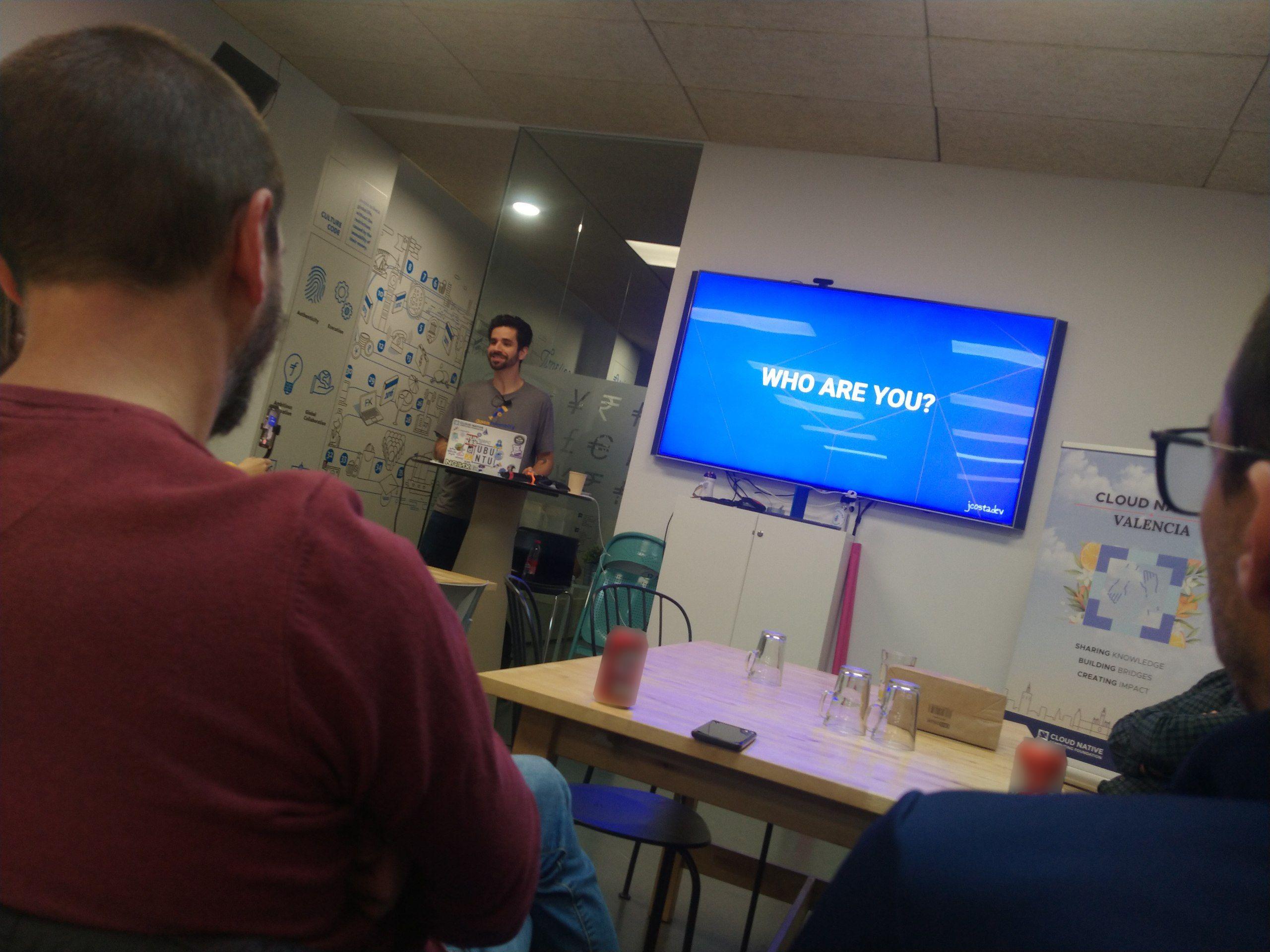
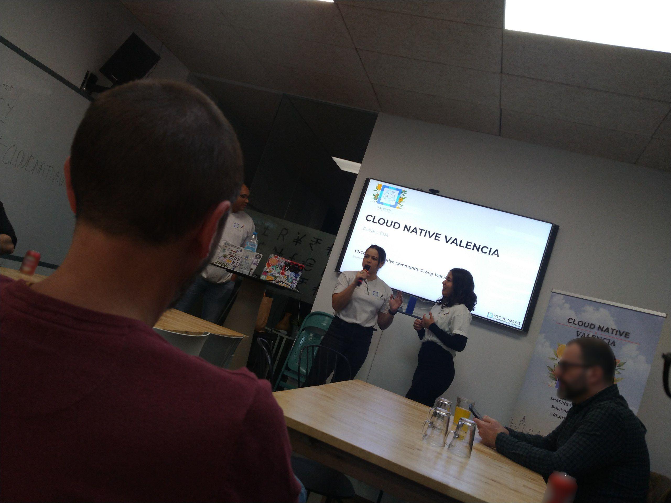
Estrenando CNCG: CNCF Valencia!
#CNCF #CLOUDNATIVEVALENCIA #FLYWIRE @julianocosta89

Durchbruch: Neurowissenschaftler haben erstmals den Schaltplan des gesamten Gehirns eines komplexen Tieres erstellt – dem der Fruchtfliege. #Gehirn #Hirnkarte #Hirnkartierung #Dosophila #FlyWire
https://www.scinexx.de/news/psychologie/erster-schaltplan-eines-gesamten-gehirns/

Tiny brain, big deal: fruit fly diagram could transform neuroscience;
Scientists took years to map 50m connections, which may lead to understanding of how wiring gives rise to behaviour
https://www.theguardian.com/science/2024/oct/02/fruit-fly-brain-connections-wiring-diagram-neuroscience
#news #neuroscience #science #brain #fruitfly #computerscience #connectomics #flywire

the whorls in that recently released cubic milimeter of human brain segmentation data might be present in drosophila.
what do you all know about these spiral, condensed neural cells? has anyone assigned tissue direction vectors to voxels of segmented data and then computed the curl of that vector field as a way to find spirals?

Does anyone know what happened with the #Flywire Android App? I don't even see it in Google Play.

The connectome of the adult female fly brain, Drosophila melanogaster:
“Neuronal wiring diagram of an adult brain” by Dorkenwald et al. 2023 https://www.biorxiv.org/content/10.1101/2023.06.27.546656v1
A project that started with Davi Bock and Wei Lee developing the TEMCA for the mouse visual cortex in Clay Reid’s lab (Bock et al. 2011 https://www.nature.com/articles/nature09802), then Bock moving to #HHMIJanelia and developing the instrument much further to deliver the #FAFB volume (Zheng et al. 2018 https://www.sciencedirect.com/science/article/pii/S0092867418307876 ), and then the Seung, Murthy and Jefferis labs automatically re-registering, segmenting and annotating the whole volume via #FlyWire and more.
Extraordinary!

If you're interested in my process on #Flywire proofreading #Drosophila, here's my run through video. This doesn't serve as a full newbie tutorial - but should give an idea of how I go through each cell I review.
Recorded a half hour run through of my #Flywire process last night, working on getting it processed now.
This was an off the cuff recording, primarily designed to walk a partnered researcher through my process, but I'm open to making more videos about things in the future.

@brembs very intrigued by how can motor neurons and vision info communicate in this operant self learning! Wondering if #TransTango or #Flywire would shed some light?
Spent the past couple of hours untangling this set of #Drosophila neurons on #Flywire which had been improperly merged by the AI due to a misalignment in the EMs.
Misalignments are one reason humans are still needed to proofread connectomes - imaging is amazing, but not perfect!
Shown here are a TmY-type, a T4, and several T5s in the optic lobe.
I go by Jay, they/she.
I am the #Editor of MMOHuts and OnRPG. I have professionally written about #VideoGames since 2008.
I am passionate about #CitizenScience - my current project is mapping the connectome of #Drosophila through #Flywire. #Biology nerd.
I am also a digital artist, but I have a separate account for that: @lithiumreflections . I also enjoy #Photography.
I do my best to be #AntiRacist. I believe in boosting voices, not replacing them.
I want to share an awesome tool that #Flywire #CitizenScience has put together. Colleague and fellow CitSci "Krzysztof_Kruk" put together a "tool" that makes identifying T4 and T5 subtypes much easier.
The link contains HS (layer 1), H1 (layer 2), and VS (layer 4) neurons. By checking the terminals of a T4/T5 neuron in relation to these, you can identify them as a,b,c,d subtypes.
You have to marvel how #Drosophila has made its own clear layering system!
@EditorSansSerif What I find amusing about the arbor morphology of this #Drosophila neuron is how much it reminds me of neurons in #Celegans!
Do you see a left-right pair of partners?
Are there more neurons within its lineage with similar morphology?
Does it target brain areas known for compact neuropils, like compartments of the protocerebral bridge?
While I'm at it, let me reach out to you wonderful #drosophila folks.
We (#Flywire citizen scientists) have come across a neuron that does not fit any types we know. Several of us have looked to see if there is an error in the trace (such as a merger) but have been unable to find any. The upper portion looks like a C-type, but it begins outside the lobula plate and goes through all three optic lobe neuropils.
Discussion link: https://discuss.flywire.ai/t/what-type-of-cell-it-is/105/317?u=azurejay
Would love insight or guidance!
Today I finished proofreading and classifying the outermost layer of somas in the #drosophila lobula plate cell rind through #Flywire, mapping over 270 cells!
I've broken down my findings of cell types here: https://discuss.flywire.ai/t/gallery-of-amusements-flywire-edition/34/255?u=azurejay
I'm so thrilled to have completed this first step in mapping the outer layer of lobula plate cells - all as part of #CitizenScience!
Now it's time to go another layer deep and repeat the process. :)
Need to share this great #Flywire render video!
From the video description:
"Gorgeous DPM neurons from the brain of a fly. These innervate the mushroom body.
Coming in at a combined length of 7.7 cm and looking like an elvish crown, these cells were originally identified in FlyWire by Markus Pleijzier from Gregory Jefferis lab.
Philipp Schlegel figured out that the interesting hollow tube structure actually contains fibers from the mushroom body. "
Client Info
Server: https://mastodon.social
Version: 2025.07
Repository: https://github.com/cyevgeniy/lmst