#Neuroimaging

2025-06-19

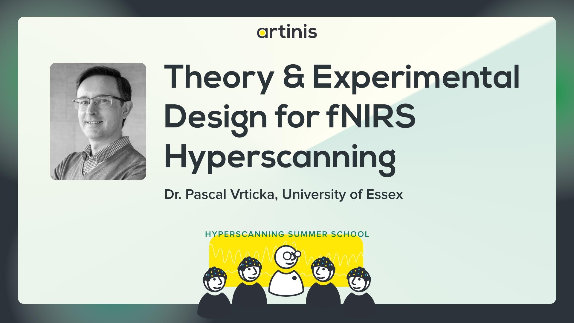
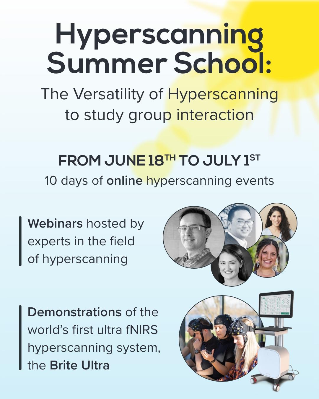
🟡Join us for today’s highlight at the Artinis #Hyperscanning Summer School!

Thursday, 19th of June
4:00–5:30 PM CEST 🌍

🎙️ Keynote 1: Dr. Pascal Vrtička ( @University of Essex )
“Theory & Experimental Design for fNIRS Hyperscanning Experiments”

This is an open platform event, meaning it’s not too late to register👉 events.teams.microsoft.com/eve

#fNIRS #EEG #neuroimaging

Matt McCormickthewtex@fosstodon.org
2025-06-19

🧠✨ Want to 3D print your own brain? Now you can—right in your browser, for free!

Try brain2print.org:

- Drag & drop your T1 MRI

- AI segmentation & mesh creation, all in-browser

- No installs, no accounts, no data leaves your computer

- Export ready-to-print STL/OBJ/MZ3 files in minutes

See it in action:
🎥 youtube.com/watch?v=_etI1yMVeg

Read the science: doi.org/10.1038/s41598-025-000

#Neuroimaging #3DPrinting #OpenSource #Privacy #MRI #Brain2Print

2025-06-18

The Hyperscanning Summer School begins today, 18th of June, with the opening talk “Introduction to fNIRS and #Hyperscanning” by Sophie Apprich from 4:30 to 5:00 PM CEST 🌍.

Join the talk last minute 👉 events.teams.microsoft.com/eve

#fNIRS #EEG #neuroimaging

2025-06-17

Functional MRI is invaluable for understanding #brain function, but findings are often of limited real-world relevance. Benjamin Becker & Feng Zhou discuss how #neuroimaging in more naturalistic environments may reveal crucial insights into human #cognition and social interactions. 🧪
plos.io/3HHcxXH

CSBJcsbj
2025-06-08

🧠 Can brain volume measurements forecast the course of multiple sclerosis?

🔗 Assessing the role of volumetric brain information in multiple sclerosis progression. Computational and Structural Biotechnology Journal, DOI: doi.org/10.1016/j.csbj.2025.05

📚 CSBJ: csbj.org/

Assessing the role of volumetric brain information in multiple sclerosis progression. Computational and Structural Biotechnology Journal, DOI: https://doi.org/10.1016/j.csbj.2025.05.003
2025-06-02

🧠 #Brain age proposed as a #biomarker to improve diagnosis and treatment for patients suffering from #neurodegenerative diseases using #DeepLearning & #BigData.

📃 Grant Proposal: doi.org/10.3897/rio.11.e156738

#neuroscience #neuroimaging

2025-05-28

The Neuromatch #ImpactScholars Program is a global initiative that provides early-career researchers with hands-on mentorship, collaborative project work, & next steps to grow as computational scientists.

🤓 Saameh Sanaaee is a cell & molecular biology graduate from Iran, currently conducting #neuroimaging research. She shares her story: linkedin.com/posts/neuromatch_

✨ Learn more about the Neuromatch Impact Scholars Program: lnkd.in/gD9ETfay

#MyNeuromatchStory #Neuromatch #Neuroscience

2025-05-27

Our latest blogpost highlights how physiological signals can provide valuable insights when properly analyzed. With some #fNIRS devices, researchers can turn physiological parameters into complementary data sources.

🔗 artinis.com/blogpost-all/non-c

#Neuroimaging #BrainResearch

Alexander Weberweberam2
2025-05-20

Using a multimodal MRI approach 🧲📊, we generated detailed semiregional brain maps of oxygen use in infants born <32 weeks GA, scanned at term-equivalent age. T1, T2, ASL, and SWI sequences were included.
🧵 2/9

Diffusion Imaging in Pythondipy
2025-05-07

🚨 Flipped b-vectors in your diffusion MRI data?
The Fiber Coherence Index (FCI) by Schilling et al. is here to help!

🔎 Detect and correct B-table orientation errors with ease.
📄 Read the paper(doi.org/10.1016/j.mri.2019.01.)
🔗 Already integrated into DIPY, CLI feature coming soon!

Get involved and explore the implementation:
👉 GitHub PR(github.com/dipy/dipy/pull/3447)

2025-05-05

'Improved Alzheimer Disease #Diagnosis With a #MachineLearning Approach and #Neuroimaging: Case Study Development' - a #Research article by JMIR Publications on #ScienceOpen: scienceopen.com/document?vid=9

2025-05-01

To protect an individual's #privacy, facial features are removed before data-sharing of human #neuroimaging data. @oesteban &co show that this procedure affects the quality ratings given by human raters but not automated image quality metrics @PLOSBiology plos.io/3Gtr3lo

An example of T1w image before and after defacing. Defacing is typically implemented by zeroing the voxels around the face (left-hand side, panel “T1-weighted”). The “Background noise” panel shows two visualizations extracted from the MRIQC visual report, in which a window is applied to select the lowest intensities and then they are inverted to enhance patterns in the background. The red arrows indicate aliases along the anterior–posterior axis—which had the lowest bandwidth in the example—produced by eye motion. These aliases are straightforward to notice in front of the ocular globes in the “Background noise” panel of the “nondefaced” image because of the absence of signal sources. This aliasing also spreads in the opposite direction, overlapping brain tissue. However, this overlap is often very hard to notice against the signal of interest within the brain. The corresponding “defaced” version of the “Background noise” panel shows how defacing eliminates valuable information for quality assessment.
CSBJcsbj
2025-04-24

🧠 Is AI ready to take the lead in brain atrophy detection?

🔗 Deep learning-based automatic segmentation of brain structures on MRI: A test-retest reproducibility analysis. Computational and Structural Biotechnology Journal, DOI: doi.org/10.1016/j.csbj.2025.04

📚 CSBJ Smart Hospital: csbj.org/smarthospital

Deep learning-based automatic segmentation of brain structures on MRI: A test-retest reproducibility analysis. Computational and Structural Biotechnology Journal, DOI: https://doi.org/10.1016/j.csbj.2025.04.007
2025-04-22

Ultrafast optical imaging captures complex neuronal activity in real time, with promise to improve understanding and treatment of conditions like Alzheimers and Parkinsons. Learn how ultrafast #NeuroImaging is shaping brain research: www.spiedigitallibra... #Neurophotonics #Neuroscience

Examples of neuro-volumetric imaging.
2025-04-22

Our latest blogpost breaks down the key advantages of fNIRS vs. fMRI. Spoiler: our #fNIRS devices meet the demands of cutting-edge clinical research, making brain imaging easy everywhere and for everyone.
🔗 bit.ly/42QgRfe

#ClinicalResearch #Neuroimaging #Neuroscience

2025-04-21

Newly developed cannula system delivers imaging agents + drugs directly for high-resolution deep tissue brain imaging. Results demonstrated for Alzheimers research - learn more: spie.org/news/pushin... #Neurophotonics #NeuroImaging #Neuroscience

2025-04-17

Next to CE certification according to #MDR (#EU 🇪🇺), the MediBrite is also registered as a medical device in the Medicines & Healthcare products Regulatory Agency (#MHRA) for its clinical use in the #UK 🇬🇧

🟢 #Neuroimaging #fNIRS #NIRS

2025-04-16

We’re thrilled to announce the official launch of the Brite Ultra, our cutting edge mass #fNIRS hyperscanning system. Join us at our Hyperscanning Summer School to discovery its full potential!

🔗 Learn more & register: artinis.com/hyperscanning-summ

#Hyperscanning #Neuroimaging #BrainResearch

Client Info

Server: https://mastodon.social
Version: 2025.04
Repository: https://github.com/cyevgeniy/lmst