
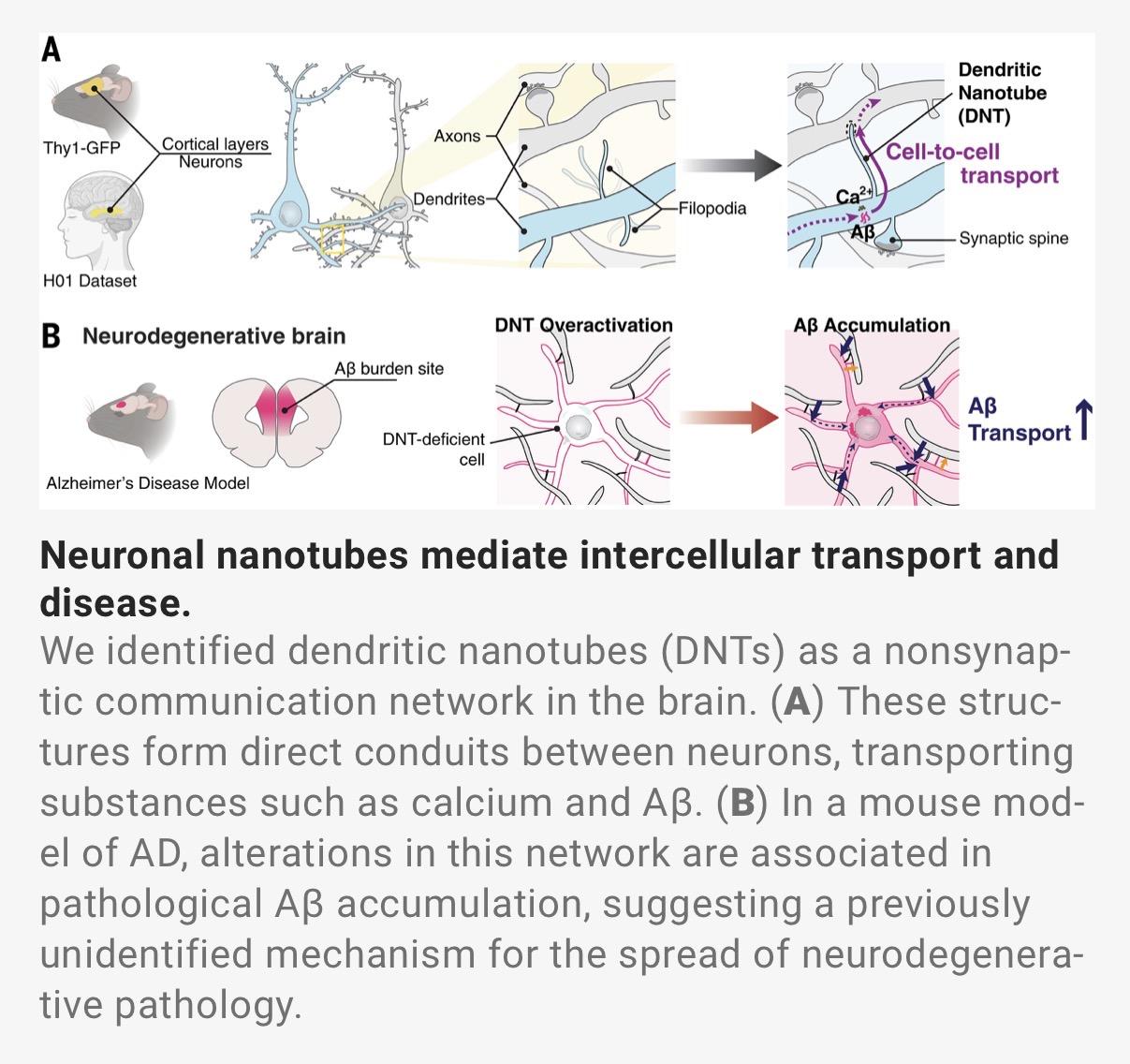
🧠 #Neurons can communicate via a hidden network of #nanotubes, new study finds. Chang et al. (2025) show that dendritic nanotubes (DNTs) are #actin-based #dendrite-to-dendrite conduits transferring Ca²⁺ and molecular cargo (incl. #Aβ), rising before plaque deposition in mouse #cortex. This overlooked pathway may expand our view of neuronal communication beyond #synapses:
#Synapses

🧠Wie Synapsen zusammenhalten 🤝
Diese Entdeckung hat ein Kölner Forschungsteam über die molekulare Architektur von Synapsen gemacht. Die Studie zeigt, dass das Protein Gephyrin im Gehirn flexible Filamente bildet und damit als wichtiger Baustein inhibitorischer Synapsen dient.
Mehr dazu▶️ https://uni.koeln/YAZEK
📰 https://www.nature.com/articles/s41467-025-63748-w
#uniköln #unicologne #Forschung #Synapsen #Gehirn #Neurologie #Research #Synapses #Brain #Neurology
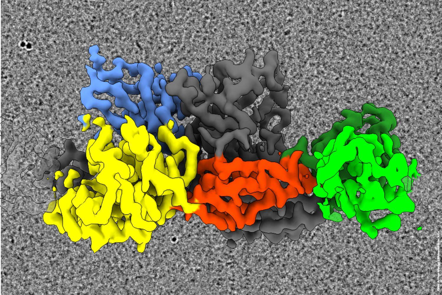
🧠 How Synapses Hold Together 🤝
This discovery was made by a Cologne-based research team studying the molecular architecture of synapses. The study shows that the protein gephrin forms flexible filaments in the brain, serving as an important building block of inhibitory synapses.
Read more ▶️ https://uni.koeln/PSVRY
📰The study was published in Nature Communications: https://www.nature.com/articles/s41467-025-63748-w
#uniköln #unicologne #Forschung #Synapsen #Gehirn #Neurologie #Research #Synapses #Brain #Neurology

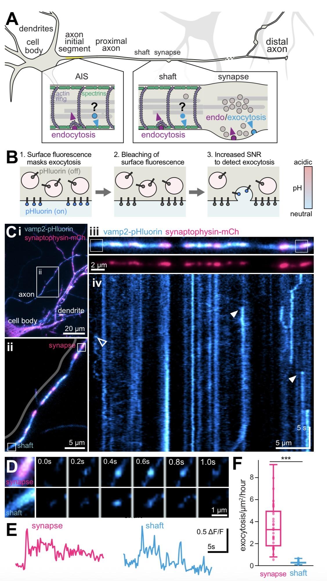
🧠 New pre-print by Wiesner et al. (2025) shows non-#synaptic #exocytosis directly from the #axon shaft, regulated by the submembrane periodic skeleton. Using #superresolution #imaging and live assays (#HiLo (VAMP2-pHluorin), #SIM, and correlative two-color #SMLM/ #STORM) they reveal that #axons can release vesicles outside classical #synapses, expanding how we understand #neuronal communication and #AxonalSignaling.

Ah, the riveting tale of psychiatry's #metaphorical #brain babble, where #neurons are poets and #synapses sing 🎤. #Nature.com suggests upgrading your #browser because, apparently, without the latest CSS, you can't truly grasp the profound wisdom of metaphorical mush 💻🔍.
https://www.nature.com/articles/s41380-025-03053-6 #psychiatry #upgrade #HackerNews #ngated

Piangere non è un sintomo.
È il modo in cui il cervello riscrive se stesso.
Ogni lacrima contiene memoria, ormoni, riorganizzazione.
Ogni abbandono lascia una traccia nelle sinapsi.
Ogni emozione profonda… ci cambia per sempre.
Articolo completo sul blog
#michiyospace #neuroplasticità #emozioni #pianto #neuroscienze #lacrime #doloreemotivo #connessioni #sinapsi #abbandono #menteecorpo #connettoma
Crying is not a symptom.
It’s how the brain rewrites itself.
Every tear contains memory, hormones, reorganization.
Every abandonment leaves a trace in the synapses.
Every deep emotion… changes us forever.
Full article on the blog
#neuroplasticity #emotions #crying #neuroscience #tears #emotionalpain #connections #synapses #abandonment #mindandbody #connectome
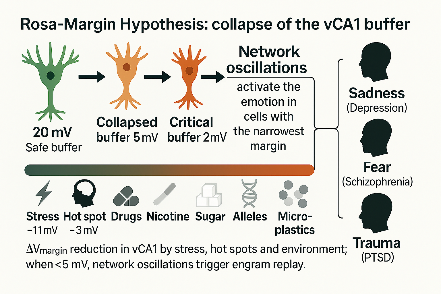
One mechanism, three disorders: When the excitability margin in vCA1 neurons falls <5 mV, network oscillations replay emotional engrams → depression, schizophrenia, PTSD.
🔗 https://doi.org/10.31219/osf.io/e4cwb_v1
#Science #Neuroscience #Neurobiology #Neurophysiology #Brain #BrainHealth #MentalHealth #Psychiatry #Schizophrenia #Depression #PTSD #Hippocampus #Memory #Synapses #CognitiveScience #NeuralNetworks #OpenScience #FediSci #Preprint #AcademicChatter #AcademicTwitter #SciComm #Researchers #Research #OSF
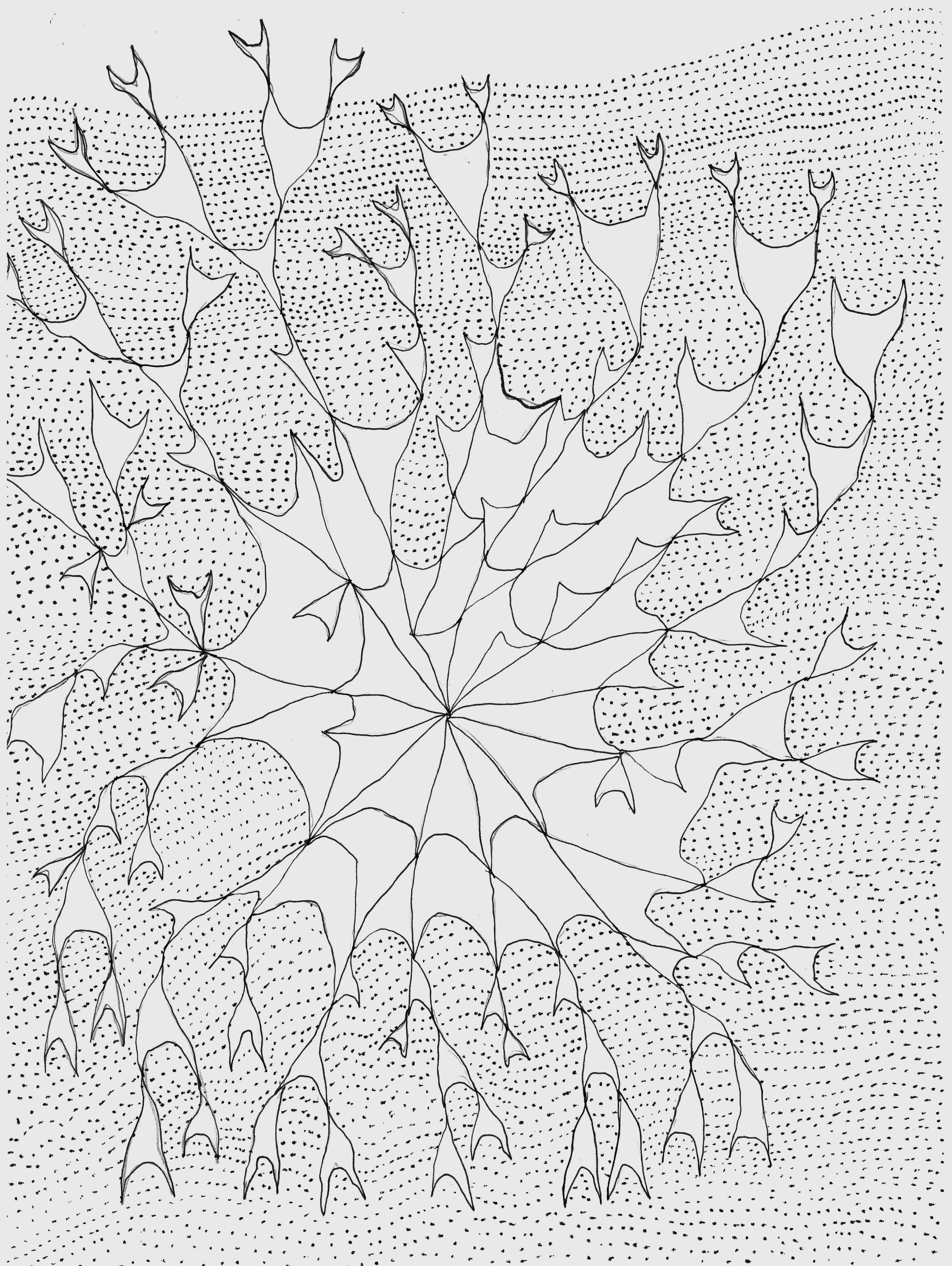
Improvisation graphique du mercredi. utilisation d'un motif se répétant en variation.
#dessin #graphisme #drawing #synapses

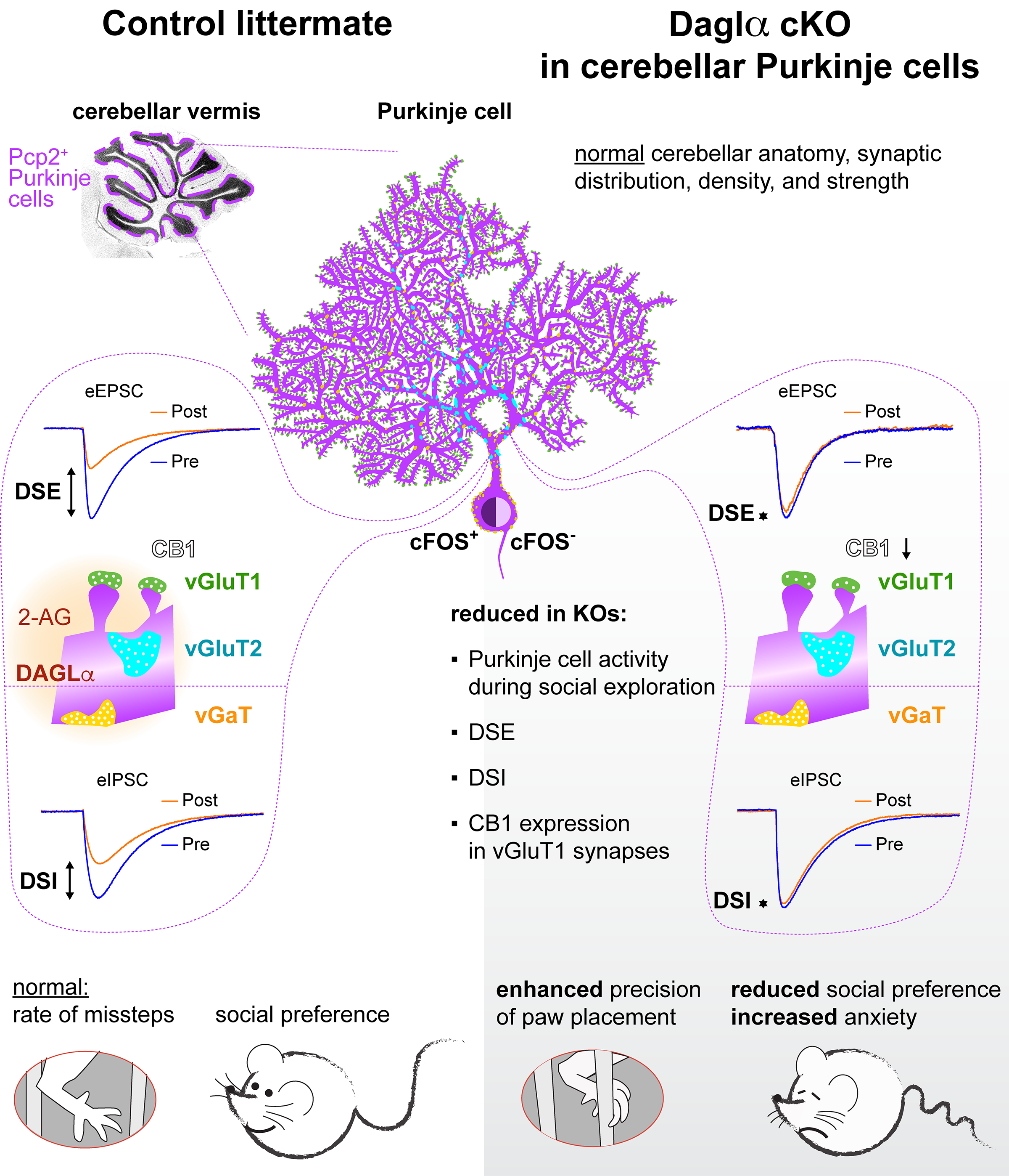
Our new research on cannabinoid signaling in cerebellar development and function is published in eNeuro.
#cannabinoid #cerebellum #Purkinje #DevelopmentalNeuroscience #endocannabinoid #indianauniversity #synapses #braindevelopment

Molecular Glue of the Brain: Cell Adhesion Molecules and Synaptic Stability
#Neuroscience #BrainScience #Synapses #NeuronalCircuits #Neuroplasticity #ExcitatoryInhibitoryBalance #DendriticSpines #ExtracellularMatrix #Interneurons #CognitiveHealth #Neurobiology #BrainDevelopment #NeuralConnections #Neuropsychiatry #BrainFunction

The #Brain stores #Memories primarily by strengthening or weakening the connections between neurons, called #Synapses.
This process, known as synaptic plasticity, allows neural circuits to be physically reshaped, forming robust pathways that encode learned information.
https://knowledgezone.co.in/posts/How-does-the-Brain-Store-Memory-678603b1a7e475eb5e304048
Exploring the Brain: A Journey Through Neuroimaging and Neural Dynamics
#BrainScience #Neuroimaging #Neuroscience #BrainHealth #Microglia #Synapses #Endocannabinoid #Neurochemistry #BrainArchitecture #Memory #Cognition #MoodRegulation #Neurotherapy #BrainConnectivity #MentalHealth #NeuroscienceExplained

Synaptic transmission is modulated by #neuropeptides in sensory systems & interneurons. This study shows that motor #synapses are also modulated by neuropeptidergic signaling, where lower ACh levels are compensated by upregulation of postsynaptic Ca2+ channels @PLOSBiology https://plos.io/4d6Ecgg
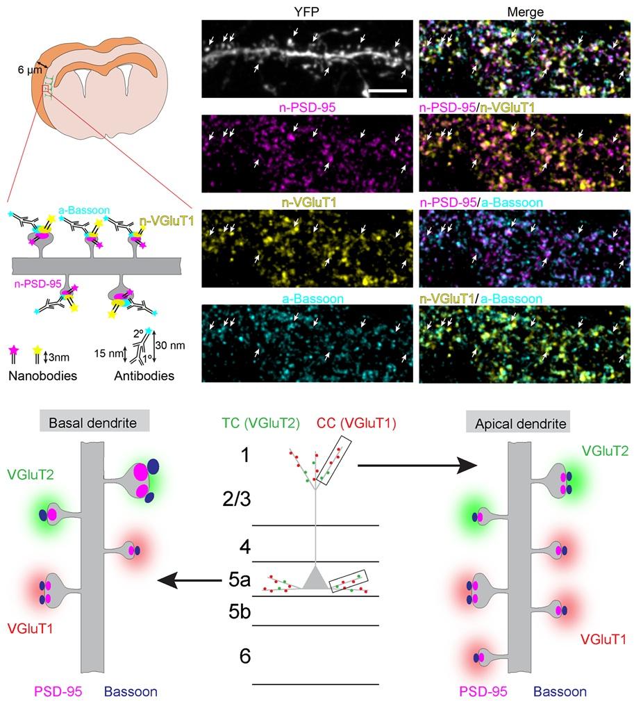
How do diverse synapses in the brain organize their nanoarchitecture? This study uses #nanobodies & STED imaging to study the nanoarchitecture of thalamocortical & corticocortical #synapses, revealing distinct principles of synaptic nano-organization in the brain @PLOSBiology https://plos.io/3FY4vcd

WRITER FUEL: Human brains take in sensory data at more than 1 billion bits per second, but only process that information at a measly 10 bits per second, new research has found.
#LimFic #LiminalFiction #WriterFuel #Writers #Authors #WritersofMastodon #StoryIdeas #HumanBrain #Synapses #Processing

Role of axo-axonic synapses among olfactory receptor neurons (ORNs) in odor decorrelation, mediated by metabotropic acetylcholine receptors:
"Nonlinear high-activity neuronal excitation enhances odor discrimination", Julia E. Manoim-Wolkovitz et al. 2025
https://www.cell.com/current-biology/fulltext/S0960-9822(25)00198-8
#neuroscience #Drosophila #AxoAxonicSynapses #synapses #olfaction
#Cellular structure without a #membrane: Researcher discusses how #synapses use #liquids to create functional separations.
#LLPS #membrane-less_organelles #neurotransmission
https://phys.org/news/2025-03-cellular-membrane-discusses-synapses-liquids.html
"The first synapse was visualized using electron microscopy in the mid–1950s, although parallel work and delicate egos make it difficult to determine with certainty the first researcher to actually see one." – Alexandra Balwit https://www.asimov.press/p/barcoding-brains
Neuroscientists are human after all.
Synapses in the fly brain learning and memory centre, the mushroom body:
"Quantitative characterization of the pattern of Brp clusters across multiple individuals revealed cell-type-dependent synapse heterogeneity and stereotypy. Furthermore, we discovered previously unidentified sub-compartmental synapse configuration and its regulation by cAMP signaling."
From:
"High-throughput synapse profiling reveals cell-type-specific spatial configurations in the fly brain", by Wu et al. (Tanimoto lab) 2025
https://www.biorxiv.org/content/10.1101/2024.12.02.626511v4
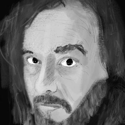
Digital painting: Ambiguous impulses for your mental cinema!
#kunst #contemporaryArt #mentalCinema #digitalPainting #virtualPainting #aiAssistedArt #saveBioDiversity #climateActionNow #systemChange #endFossilFuels #makeArtNotWar #noAFD #NoRacism #NoFascism #humanrights #humanDignity #Нетвойны #anatomy #brain #neocortex #neurosciences #neurobiology #cognition #perception #emotion #synapses #astronauts #spaceSuit #spaceTravel #tightRopeWalking #balancing #berlin #reichstag #government #capital
Client Info
Server: https://mastodon.social
Version: 2025.07
Repository: https://github.com/cyevgeniy/lmst