Allan Carrillo-Baltodano

Interested in marine embryos and larvae; tico 🇨🇷 in London 🏴󠁧󠁢󠁥󠁮󠁧󠁿; #embryo2019 #LatinoEnCualquierParte he/him/his

Allan Carrillo-Baltodano boosted:
2023-09-12

Meet the lab of Chema Martín-Durán @Chema_MD, based at Queen Mary University of London, UK.

The lab studies segmented worms with spiral cleavage to understand how development is controlled and evolves to generate new phenotypes #EvoDevo

thenode.biologists.com/lab-mee

Allan Carrillo-Baltodano boosted:

For our #MidweekMovie, we are recommending that you catch up with the #DevPres talks on @the_node. The speakers, Hannah Gruner, Seth Donoughe & @allan_littlecar all use microscopy to address research questions in #evodevo.

thenode.biologists.com/develop

#DevBio

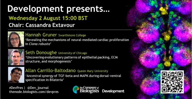
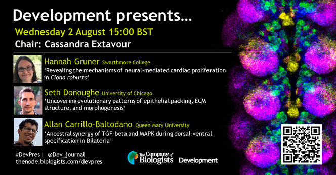
Development presents...  poster Wednesday 2 August 15:00 BST  Chair: Cassandra Extavour  Hannah Gruner (Swarthmore College) ‘Revealing the mechanisms of neural-mediated cardiac proliferation in Ciona robusta’ 
Seth Donoughe (University of Chicago)  ‘Uncovering evolutionary patterns of epithelial packing, ECM structure, and morphogenesis’ 
Allan Carrillo-Baltodano (Queen Mary University) ‘Ancestral synergy of TGF-beta and MAPK during dorsal-ventral specification in Bilateria’  #DevPres | @Dev_journal  thenode. biologists.com/devpres
Allan Carrillo-Baltodano boosted:
2023-08-08

Genomic Analysis of a Scale Worm Provides Insights into Its Adaptation to Deep-Sea Hydrothermal Vents doi.org/10.1093/gbe/evad125 #science #evolution #biology #genome #evolgen_paper

A photo of Branchipolynoe longqiensis as well as a bubble blot on the mean GC content, sequencing coverage and species hit of the genomic assembly
Allan Carrillo-Baltodano boosted:
2023-08-07

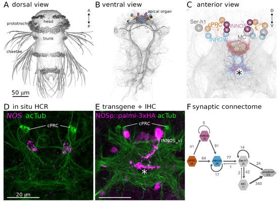
We published a new preprint on nitric oxide (NO) signalling in #Platynereis. In the paper Kei Jokura et al. described how NO is required for UV-avoidance behaviour by mediating retrograde signalling to brain ciliary photoreceptors.
biorxiv.org/content/10.1101/20
#neuroscience #animals #science

Multi-panel figure: Identification of NOS-expressing interneurons (INNOS) within the cPRC circuit. (A) Scanning electron microscopy image of a three-day-old Platynereis larva. (B, C) Volume rendering of the neuron types (cPRC, INNOS, INRGWa, Ser-h1 and MC) in the cPRC circuit reconstructed from a whole-body transmission electron microscopy volume of a three-day-old larva. Neurite skeletons are shown with cell-body positions represented by spheres. Projections of all neurons in the body are shown in grey to highlight the neuropils. The outline of the yolk is also indicated in grey. In B, nuclei positions of the prototroch head ciliary band are shown as grey spheres. (D) Expression of the NOS gene detected by in situ HCR (magenta) in a two-day-old larva (anterior view). Antibody staining for acetylated α-tubulin (acTub: green) highlights cPRC cilia and the neuropil. (E) Expression of a NOS reporter (NOSp::palmi-3xHA; magenta) labelled with an anti-HA antibody in a two-day-old larva (anterior view). Antibody staining for acetylated α-tubulin (acTub; green) highlights cPRC cilia and the neuropil. (F) Synaptic wiring diagram of the cPRC circuit.
Allan Carrillo-Baltodanoallan_littlecar@ecoevo.social
2023-07-14

🚨The Martín-Durán Lab is recruiting a PostDoc and a PhD to investigate how larvae evolved! Supported by Leverhulme Trust at Queen Mary University of London, join a terrific group of people to investigate fascinating marine 🪱🪱!

PhD ad: alturl.com/i64ue
DM/e-mail (chema.martin@qmul.ac.uk) for more info!

Allan Carrillo-Baltodano boosted:
Gaspar Jekelyjekely@qoto.org
2023-07-04

In our new preprint we characterise neuropeptide signalling in the cnidarian #Nematostella.
biorxiv.org/content/10.1101/20
Daniel Thiel and Luis Yanez-Guerra screened 64 neuropeptides against 161 G-protein coupled receptors (#GPCR) and found activating peptide ligands for 31 receptors.
#cnidaria #neuropeptide #evolution #neuroscience

Allan Carrillo-Baltodano boosted:
2023-06-30

Last, but by no means least, at #GRCdevbio23 is @Honghao_Song from @redmakeda's lab at @Harvard

Together, the Extavour lab investigates the evolution of the reproductive system.

@redmakeda is also a @Dev_journal Editor and Chair of our next #DevPres webinar on 2 August.

Take a look at the #DevPres programme of speakers on @the_node
thenode.biologists.com/devpres

Allan Carrillo-Baltodano boosted:
Bruno C. Vellutinibruvellu
2023-06-08

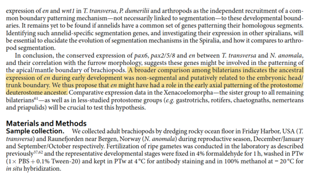
@albertcardona Nice work. I reached a similar conclusion when looking at engrailed expression across metazoans. From doi.org/10.1038/srep32387

But I don't think the conserved genetic boundary suggests the homology of brains. To me, it supports the homology of a head-trunk distinction in early development.

The brain of protostomes and deuterostomes could well have evolved independently using this common genetic basis.

A broader comparison among bilaterians indicates the ancestral expression of engrailed during early development was non-segmental and putatively related to the embryonic head/trunk boundary. We thus propose that engrailed might have had a role in the early axial patterning of the protostome/deuterostome ancestor.
Allan Carrillo-Baltodano boosted:
2023-05-03

Spatial transcriptomics sheds light on the #echinoderm body plan!

#preLight from Rodrigo Senovilla-Ganzo.

Rodrigo covers a #preprint from #lab_lowe showing that the bilaterian antero-lateral axis shifts to medio-lateral in the #ectoderm of adult echinoderms.

👉 prelights.biologists.com/highl

Ambulacral-anterior model of echinoderm body plan evolution.
Allan Carrillo-Baltodano boosted:
Alberto Perez-Posadaapposada@genomic.social
2023-04-27

My first contribution/collaboration in the lab of Jordi Solana is out!

biorxiv.org/content/10.1101/20

First cell type atlas of the annelid Pristina leidyi uncovering a bunch old and new cell types, generating networks of gene coexpression, and identifying piwi-positive cells as pluripotent lineage giving rise of all cell types.

It was so fun to develop and learn graph methods for this work. Congrats to all the authors!

#academics #singlecell #annelids #celltypes

Allan Carrillo-Baltodano boosted:
2023-04-24

Thrilled to see our latest work highlighted on the cover of Science 🤩. Our discoveries challenge our understanding of nervous systems and their evolution. Link to the paper here:

science.org/doi/10.1126/scienc

Allan Carrillo-Baltodanoallan_littlecar@ecoevo.social
2023-04-19

In a month I'll be heading to my alma mater University of Costa Rica to present some of our work on axial specification in annelids 🪱

En un mes estaré presentando nuestro trabajo en especificación de ejes en anélidos en mi alma mater Universidad de Costa Rica

#EvoDevo #WormWednesday #CienciaCriolla

Presentation title for lecture at University of Costa Rica
Allan Carrillo-Baltodano boosted:
2023-04-18

We are really excited to share the news that for the first time, EuroEvoDevo will have five satellite meetings @EED2024 Helsinki! Endless #EvoDevo most beautiful!
Save the dates:
📅June 24 & 25, 2024: Satellites
📅June 25 – 28, 2024: Main EED Meeting

Thanks to the organizers! More info soon on euroevodevo2024.fi

Allan Carrillo-Baltodanoallan_littlecar@ecoevo.social
2023-04-14

@Chema_MD presenting our work at the Gene Regulatory Networks meeting #EvoDevo #GRN2023

Allan Carrillo-Baltodanoallan_littlecar@ecoevo.social
2023-04-08

Thanks to Bernard and Sandie Degnan for including our annelid life cycle paper in their spotlight for Trends in Genetics

"How larvae and life cycles evolve"

cell.com/trends/genetics/fullt

Good that our work keeps on making a contribution to the community

#EvoDevo #Annelids

Allan Carrillo-Baltodano boosted:
Bruno C. Vellutinibruvellu
2023-04-03

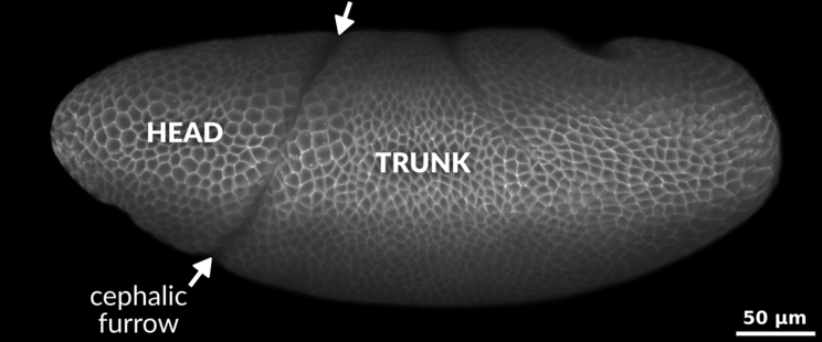
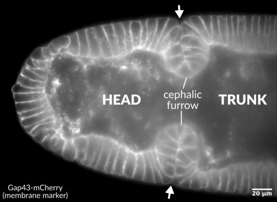
I'm excited to announce that our cephalic furrow paper is out! We investigated the function of this enigmatic invagination using live imaging and modeling and found that it plays a mechanical role during fly gastrulation biorxiv.org/content/10.1101/20

Lateral view of a Drosophila embryo (fruit fly) rendered in 3D showing the cell membranes. The head (to the left) and the trunk (to the right) are annotated. In between there is a large invagination known as the cephalic furrow. Arrows delimit the ventral and dorsal limits of the cephalic furrow. A scale bar on the right corner reads 50µm. The embryo has about 500µm in length.An optical slice from a Drosophila embryo (fruit fly) showing the invaginations of the cephalic furrow on the left and right sides.
Allan Carrillo-Baltodano boosted:
2023-03-30

We made a book! If you like weird and wonderful invert eyes, check it out:
link.springer.com/book/10.1007

Featuring awesome contributions form a load of great researchers.

Allan Carrillo-Baltodanoallan_littlecar@ecoevo.social
2023-03-24

#FluorescentFriday

Some antibodies arrived a few weeks ago, so we tested in Capitella. Looking forward to testing them in Owenia 🪱

Allan Carrillo-Baltodano boosted:
James Briscoejamesbriscoe@mas.to
2023-03-24

Very excited to see the launch of the 1st cohort of @Dev_journal Pathway to Independence (PI) Fellows

8 postdocs we'll follow & support as they navigate the academic job market

You'll be hearing more from them & about the PI programme in the near future

journals.biologists.com/dev/pa

Allan Carrillo-Baltodanoallan_littlecar@ecoevo.social
2023-03-15

This #WormWednesday we welcomed a box full of possibilities 💪🏼

Owenia is back 🪱!!!, plus some extra spiralian friends 🐌

Thanks to the Roscoff Marine Station for the package 🏴󠁧󠁢󠁥󠁮󠁧󠁿🤝🏼🇫🇷

Client Info

Server: https://mastodon.social
Version: 2025.04
Repository: https://github.com/cyevgeniy/lmst