
🎉 Behold the tale of the electron microscope! A century of progress boiled down to "now you can see really tiny stuff." 📏🔬 Meanwhile, your browser still can't play audio—it's like pioneering 1920s technology on a 2025 device. 🚀🦖
https://www.asimov.press/p/electron-microscope #electronmicroscope #technologyprogress #tinyworld #browserissues #2025innovation #HackerNews #ngated
#electronmicroscope


💌 Our first Valentine comes from the #EMGlossary 💌
Thanks to our amazing community 👨🔬 👩🔬 v2.0.0 of our OWL artefact is now published 😍
Love knows no boundaries and neither should data. The electron microscopy glossary ensures that data crosses labs and disciplines effortlessly. #Interoperability
🌐 https://emglossary.helmholtz-metadaten.de
#electronmicroscopy #electronmicroscope
#LoveData25
#LoveDataWeek
#DataMatters Hashtag
#HMC @helmholtz_hmc


Donating old #instruments to labs that can’t afford to buy new sounds like a nice idea, but the practicalities are often difficult (for example with an #ElectronMicroscope a colleague got, not noticing the liquid mercury cap was open and the box upside-down). https://cen.acs.org/business/instrumentation/happens-old-scientific-instruments/102/i18?utm_source=Live+Audience&utm_campaign=c11700db61-nature-briefing-daily-20240625&utm_medium=email&utm_term=0_b27a691814-c11700db61-50179668


Groundbreaking Images Reveal the Human Brain at Nanoscale Resolution https://petapixel.com/2024/05/13/groundbreaking-images-reveal-the-human-brain-at-nanoscale-resolution/ #electronmicroscope #neuroscience #microscope #microscopy #Spotlight #science #brain #News

Exploring the Unseen Universe: The Marvels of the Electron Microscope
#ElectronMicroscope, #Microscope, #ScanningElectronMicroscope, #SEM, #TEM, #TransmissionElectronMicroscope #Instrumentation Introduction The electron microscope stands as one of the most remarkable inventions in the history of science and technology. It…. Medical Microbiology & Recombinant DNA Technology (RDT) Labs | Read More -

Eva Weig, Professor for Nano and #QuantumSensors, and her team are producing vibrating nanostrings large enough to be seen under an #electronmicroscope. They could become fundamental components of a new #quantumtechnology: http://go.tum.de/091531
@MCQST
📷M.Jooss

Time flies by. ⌛ One year ago, the DGE Young Microscopists (yDGE) were founded. In the first year, we set up a website www.ydge.de, 🌐 hosted our first social events and planned the first yDGE conference contribution 👔 (check out https://www.microscopy-conference.de/2/programme/scientific-programme WS 3).
#mc2023 #microscopyconference #microscopy #microscope #electronmicroscopy #electronmicroscope #science #imaging
The stuff he's making may be obsolete, but rediscovering obsolete technology may be a path to improving current technology.
It's also pretty cool that he bought a broken $250k electron microscope for $1000 and repaired it.
https://www.wired.com/story/22-year-old-builds-chips-parents-garage/
#semiconductors #chips #ElectronMicroscope
More old #electronics equipment from my Ebay-enabled home lab. #NixieTubes! This 1969 #Keithley 615 #electrometer has a discrete MOSFET front-end and a dual-slope A/D made of gate-level RTL chips. The rear-panel parallel output is labeled PRINTER, because that is all you were likely to do with it in 1969. I once used it to measure the beam current of a Philips #electronMicroscope. Here it is getting the right answer for a #Victoreen 1 GΩ resistor.
Butterfly eye, x1800
Focus drifted right before I hit start scan but I didn't notice until part way through. So it goes with scanning microscopy.
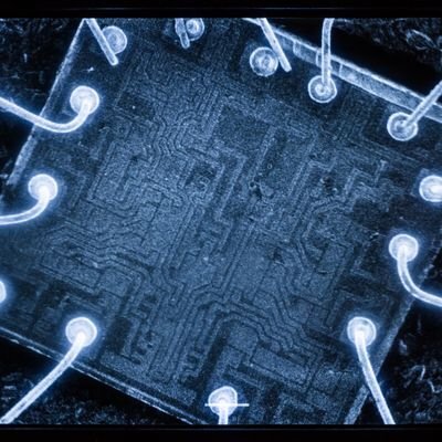
A few of our SEMs have these external scan generators/frame grabbers that ~worked. All of the scans from the 35C before this year were captured on one (including this scan of an ICE40 FPGA).
Problem is they use a proprietary fiber-optic PCI card. The software is windows XP only and infuriating to use.
I traced out the PCB for the X/Y sweep DACs + buffer/offset circuits, then captured that in KiCad.
Seems like a great place to drop in an ICE40.

Microscopy Hack Chat with Zachary Tong
Join us on Wednesday, June 23 at noon Pacific for the Microscopy Hack Chat with Zachary Tong!
There was a time when electronics was very much a hobby that existed in the macroscopic world. Vacuum tubes, wire-wound resistors, and big capacitors were all mounted on terminal strips and mounted in a heavy chassis or enclosure, and interfacing with everything from components to tools was more an exercise in gross motor skills than fine. Even as we started to shrink components down to silicon chips, the packages we put them in were still large enough to handle and see easily. It's only comparatively recently that everything has started to push the ludicrous end of the scale, with components and processes suitable only for microscopic manipulation, but that's pretty much where we are now, and things are only likely to get smaller as time goes on.
The microscopic world is a fascinating one, and the tools and techniques to explore it are often complex. That doesn't mean microscopy is out of the wheelhouse of the average hacker, though. Zachary Tong, proprietor of the delightfully eclectic Breaking Taps channel on YouTube, has been working in the microscopic realm a lot lately. We've featured his laser scanning confocal microscope recently, as well as his latest foray into atomic force microscopy. In the past he has also made DIY acrylic lenses, and he has even tried his hand at micromachining glass with lasers.
Zach is pretty comfortable working in and around the microscopic realm, and he'll stop by the Hack Chat to share what he's been up to down there. We'll talk about all the cool stuff going on in Zach's lab, and see what else he has in store for us.
Our Hack Chats are live community events in the Hackaday.io Hack Chat group messaging. This week we’ll be sitting down on Wednesday, June 23 at 12:00 PM Pacific time. If time zones have you tied up, we have a handy time zone converter.
Click that speech bubble to the right, and you’ll be taken directly to the Hack Chat group on Hackaday.io. You don’t have to wait until Wednesday; join whenever you want and you can see what the community is talking about.
#hackadaycolumns #atomicforce #confocal #electronmicroscope #hackchat #micron #microscope #microscopy

This is the Highest-Ever Resolution Photo of Atoms
In 2018, Cornell researchers built a high-powered detector that set a world record for the highest resolution state of the art electron microscope which, at the time, tripled the previous resolution it could capture. Now, they've beaten their own record by a factor of two.
Cornell University's David Nutt writes that as successful as the first approach led by David Muller -- the Samuel B. Eckert Professor of Engineering -- in 2018 was, it was flawed. It only worked with "ultrathin samples that were a few atoms thick." Any thicker, and the electrons would scatter in ways that could not be disentangled.
However, on March 20, 2021, Muller again led a team that has beaten its own record with what is described as an electron microscope pixel array detector (EMPAD) that has an even more sophisticated 3D reconstruction algorithms and whose resolution is so finely tuned that the blurring visible in the finished image is due only to the motion of the atoms themselves.
The resulting photo (above) depicts an electron ptychographic reconstruction of a praseodymium orthoscandate (PrScO3) crystal, zoomed in 100 million times. The results of this experiment have been published in a full report here.
“This doesn’t just set a new record,” Muller says. “It’s reached a regime which is effectively going to be an ultimate limit for resolution. We basically can now figure out where the atoms are in a very easy way. This opens up a whole lot of new measurement possibilities of things we’ve wanted to do for a very long time. It also solves a long-standing problem -- undoing the multiple scattering of the beam in the sample, which Hans Bethe laid out in 1928 -- that has blocked us from doing this in the past.”
The microscope captures images using a computational method of microscopic imaging called Ptychography. It generates images by processing multiple coherent interference patterns that have been scattered from a particular object and looks for changes in the overlapping regions. By seeing how the pattern changes, the researchers were able to compute the shape of the object that caused the pattern.
“With these new algorithms, we’re now able to correct for all the blurring of our microscope to the point that the largest blurring factor we have left is the fact that the atoms themselves are wobbling, because that’s what happens to atoms at finite temperature,” Muller continues. “When we talk about temperature, what we’re actually measuring is the average speed of how much the atoms are jiggling.”
It is possible that the researchers could again beat their own world record by using material that is made up of heavier atoms, as they wobble less, or by cooling down the sample which also reduces the movement of atoms. It is theorized that atoms stop moving at absolute zero, but it has yet been impossible to reach that temperature. As a result, any lowered temperature that the researchers could reach may not reduce the movement of the atoms by a noticeable amount.
“We want to apply this to everything we do,” Muller says. “Until now, we’ve all been wearing really bad glasses. And now we actually have a really good pair. Why wouldn’t you want to take off the old glasses, put on the new ones, and use them all the time?”
Photo credits: Header image by Cornell University.
#news #technology #atom #cornelluniversity #electronmicroscope #electrons #engineering #photoofatom #ptychograph #worldrecord

"The Vast Viral World: What We Know (And Don't Know)", Nautilus (https://nautil.us/issue/99/universality/the-vast-viral-world-what-we-know-and-dont-know).
#Virus #Biology #Life #Microscope #ElectronMicroscope #Virology #Cell #Disease #Pathogen #Symbiosis #DNA #RNA #Science

Client Info
Server: https://mastodon.social
Version: 2025.07
Repository: https://github.com/cyevgeniy/lmst