
For #fluorescencefriday I just thought these neurons in the lateral geniculate nucleus were pretty. #sciart #microscopy #confocal
#confocal

Escurinho do confocal
Passando um tempo no escurinho do confocal.—
URL: https://brunovellutini.com/posts/escurinho-do-confocal/

A new study introduces two yellow #fluorescent proteins—mGold2s and mGold2t—that are significantly more #photostable than commonly used alternatives such as mVenus and mCitrine. Using these new #YFPs, researchers were able to extend imaging durations across diverse modalities including widefield, total internal reflection fluorescence (#TIRF), super-resolution, single-molecule, and laser-scanning #confocal microscopy. Learn more: https://ilphotonics.com/introducing-two-new-yellow-fluorescent-proteins/

#MSLASpheroidStamp: #OpenSource 3D #cell #spheroids for everyone:
-#3Dprinted molds/stamps
-time-lapse #confocal #microscopy of spheroids
-#cytotoxicity screens
Preprint: https://doi.org/10.1101/2024.05.12.593682
GitHub: https://github.com/arteys/MSLASpheroidStamp
#DIYbio #lab #instruments #MSLA #3Dprinting #bioprinting #imaging


Plantaardige zeefdruk van rode kool 🌱🌿
In opdracht van gr8 agency ben ik voor Regio West-Brabant aan de slag gegaan met een 100% plantaardige zeefdruk. Ik heb zelf roze inkt weten te maken van lokale rode kool en met een chemische reactie tussen de kleurstof en baking soda maakte ik een gradient. Het papier is gemaakt met tomatenplant. Het was een uitdaging om mijn werk volledig duurzaam te maken met veel obstakels gaandeweg maar met een uniek eindproduct.
-
-
-
-
-
-
-
-
#bioart #art #sciart #biology #contemporaryart #science #nature #scienceart #biodesign #artwork #artist #artandscience #research #microscopy #design #biomaterials #microscope #cellbiology #microbiology #biotechnology #microscopyart #drawing #digitalart #cells #science #artscience #biotech #cell #confocal


A research team developed a budget-friendly method for adapting confocal microscopes for Total Internal Reflection Fluorescence (#TIRF) microscopy. The paper presents “a simple and cost-effective method to build a #prism-based TIRF setup on a laser scanning confocal microscope. The setup is affordable, and users can use both #confocal and TIRF modes in the same setup with the same light source and optical components.” Learn more: https://ilphotonics.com/inexpensive-high-resolution-single-molecule-microscopy/
Drain fly embryo on Nikon Small World
Nikon Small World has announced the winners of this year’s photomicrography competition. My image of a drain fly embryo made it to the Images of Distinction category!
Gene expression patterns in a drain fly embryo (Clogmia albipunctata) with an open eggshell. See on Nikon Small World.—
URL: https://brunovellutini.com/posts/drain-fly-embryo-nikon-small-world/
#clogmiaAlbipunctata #confocal #diptera #embryo #image #microscopy #molecular #scienceOutreach


Interesting new #preprint about #tissueclearing and #lightsheet #microscopy in mouse ovaries. Complete with #napari-based deep-learning pipeline and detailed build instructions for 3D-printed sample chambers for solvent-cleared samples, so they can be viewed under a #confocal!
OoCount: A machine-learning based approach to mouse ovarian follicle counting and classification
Folts et al., preprint at biorxiv 2024
https://www.biorxiv.org/content/10.1101/2024.05.13.593993v1

The pipetting marathon I went through the other day was worth it—the experiment worked! Now I get to image some exciting and colorful samples in the confocal microscope, and experience that thrill of seeing something for the first time, once again 🌈🔬
Confocal microscope software interface showing a slice of a multichannel image stack with gene expression patterns in a fly embryo.
Ready for the new year: new #NSPARC detector for image scanning #microscopy is installed on the #Nikon #AX MP #multiphoton #microscope @ #MIC in #Muenster. Waiting for high-resolution multiphoton images.

Also including an interesting discussion about the feasibility of measuring photostability at all in a point-scanning system, beginning with the following curious sentence:
"Shaner et al., who belonged to the laboratory of the late Dr. Roger Tsien, pioneered and successfully standardized the quantification of photostability in the field of FP technology. "
Hello everyone,
Just moved my profile to this server - this is a good chance for a late introduction.
Interested in microscopy, bioimaging, confocal, superresolution microscopy, molecular motors and biological topics in general.
#microscopy #confocal #superresolution #molecularmotors #ruhrpott #introduction

Cardiomyocyte orientation recovery at micrometer scale reveals long-axis fiber continuum in heart walls
A new method that combines #confocal #microscopy with computer vision provides unprecedented spatial resolution of #cardiomyocyte geometry along the mouse heart wall.
Minhajuddin Sirajuddin, Kaleem Siddiqi et al

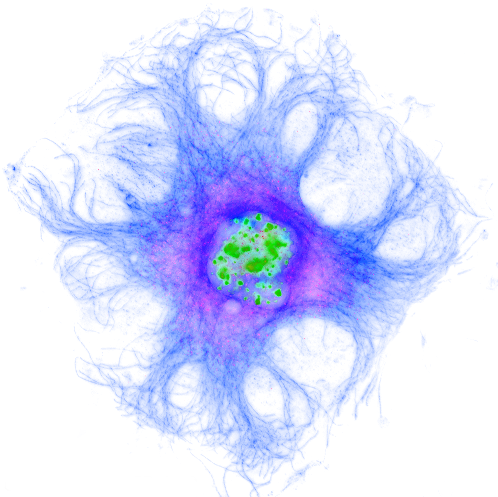
#throwbackthursday Here is an old image I took many moons ago.
#confocal #microscopy image of #MouseFibroblast cell with multiple nuclei. False coloured and inverted to be on a white background. Blue microtubules, green nuclei

RT: @svi_huygens@twitter.com
#microscopists, want to get a better overview of microscopy image #FileFormats, bit depth, scaling, image #metadata, dimensions, and more?
Join our webinar Feb 23 and explore what formats e.g. #OMETIFF:

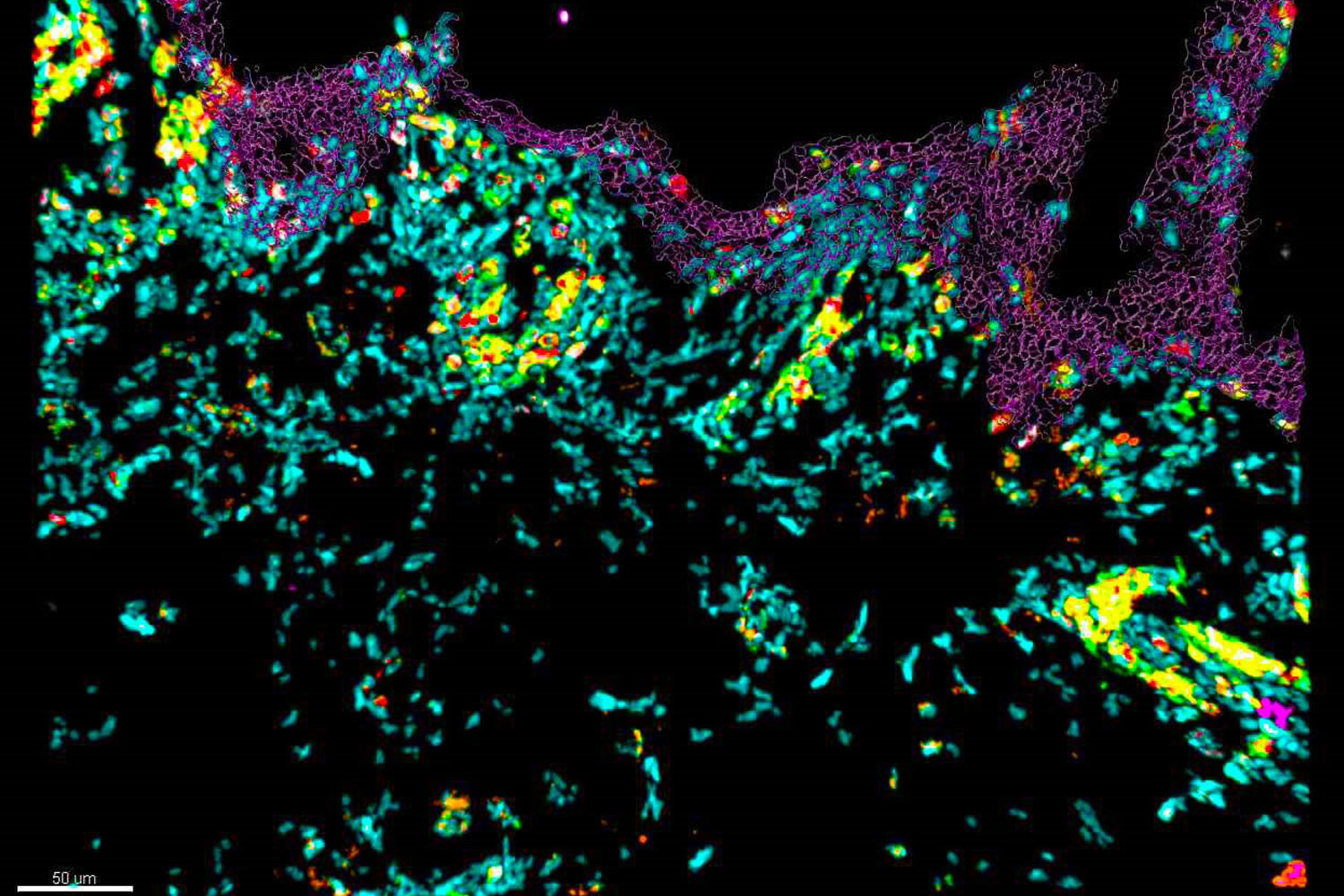
🔶 What are the additional insights you can gain from imaging tissue sample in 10 colors?
🔶 Follow our on-demand webinar with Dr. Nicolas Gaudenzio and discover how to perform 10-color imaging using a confocal microscope. See how this allows to assess the skin immune landscape.

Jan-Lukas Førde et al. present a new #software tool for automatic #cell segmentation of fluorescent #cancer cells in #zebrafish larvae:
#Imaging #Biology #BiologyOpen #Science #AcademicMastodon #Confocal #Segmentation

The new #nikon #NSPARC #AX #Confocal #microscope uses #ism image scanning microscopy and a spatial array detector to enable high sensitive #superresolution microscopy with a point scanning confocal.

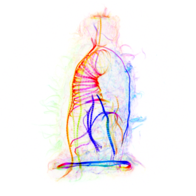
Tim Salditt (http://twitter.com/SaldittLab, substituting Jasper Frohn): #Multiscale #Xray Phase Contrast #Tomography at #GINIX/P10: Concepts, Implementation and Applications
- fantastic collaboration with the P10 team
- directly comparing #STED to #Minflux; old #confocal? not worth mentioning 😂
- overview tomo, then zoom-in
- tumorous human pancreatic tissue #biopsy: quantify the #tumor type
- from electron density to metrics, quantify fibres etc.
- #hippocampus patho punch, #Alzheimer sample
Pui-Ying (Penny) Lam imaged adult #Danionella cerebrum in vivo for the first time using standard #confocal microscopy in 2022!
Client Info
Server: https://mastodon.social
Version: 2025.07
Repository: https://github.com/cyevgeniy/lmst