#MicroscopyMonday

2025-05-21

:-( i came here hoping of #science days like back then on #twitter with #fluorescenceFriday and #MicroscopyMonday .

Instead im choking on #fascism in the world and #genocide .

there was a post some time ago about that online protest 1. doesnt work and 2. paralyzes you, giving you the illusion of being active.

i guess i pretty much failed that.
2025-02-03

Whole-mouse imaging the hard way: Clearing, then slicing + block-face imaging with beam-scan / sensor scan-line synchronization and continuous stage movement in an oblique imaging setup (ViSOR).
This time, they scaled it up to whole-mouse and used pre-cleared mouse bodies (ARCHmap-blockface-ViSOR):

High-speed mapping of whole-mouse peripheral nerves at subcellular resolution
Shi et al., preprint at biorxiv 2025
doi.org/10.1101/2025.01.22.632

#neuroscience #lightsheet #microscopy #MicroscopyMonday

Figure 1. ARCHmap-blockface-VISoR approach for high-throughput mapping of whole mouse body at micron resolution

(A) ARCHmap-blockface-VISoR pipeline for whole-mouse imaging.

(B) Bright-field view of a cleared 2-month-old vGAT-Cre;Ai14 mouse and 400-μm-thick sections collected during blockface imaging.

(C) 3D view of a ∼600-μm-thick thoracic volume imaged from a blockface imaging section.

(C1-C6) Representative high-resolution images showing maximum intensity projection of 20-μm-thick stacks in (C) at depths of 100 μm (C1, zoomed-in view in C4), 300 μm (C2, zoomed-in view in C5), and 600 μm (C3, zoomed-in view in C6).

(D, D1) Blockface-VISoR setup. (D1) Magnification of the imaging region in (D).

(E) Cross section of imaging and sectioning modules of the blockface-VISoR setup.

(F) Schematic showing overlapped imaging data (pink) of ∼200-µm thickness between 2 contiguous imaging sections.

(G) Comparison of 3D intersection alignment by natural coordinates (G1) and a custom 3D reconstruction method (G2). Images of vessel fluorescence are the maximum intensity y-projection of 20-µm-thick sections in the upper section (red) and the lower section (green).

(H) 3D view of nerve and vasculature in a whole adult Thy1-EGFP mouse. Red hot (black-red-yellow-light yellow spectrum), LEL-DL649; cyan hot (black-blue-cyan-white spectrum), Thy1-EGFP; white surface, whole mouse.
2025-02-03

Here's a #MicroscopyMonday segmentation of some neurons from the optic lobe, or visual processing area of the #Drosophila fruit fly's brain. The #connectome for the full optic lobe was released in 2024 by #HHMIJanelia, Google, and the University of Cambridge, with support from the Wellcome Trust.

Dense arbors from neurons on one side of a fruit fly's head.
2025-01-27

This segmentation of #MicroscopyMonday data reconstructs neurons of the "MANC", or male adult nerve cord, from the #Drosophila fruit fly. The nerve cord is like a spinal cord and connects the brain and muscles. #HHMIJanelia, Google, and the University of Cambridge released this work in 2023.

Dense arbors from neurons in a fruit fly's nerve cord.
2025-01-13

A use of #MicroscopyMonday data is making segmentations. Here's one, of #Drosophila protocerebral bridge and nodulus neurons, from the 2020 Hemibrain by #HHMIJanelia and Google. Michał Januszewski and Viren Jain's team made an initial segmentation with #AI flood-fill algorithms...

Long fibers from neurons at the center of a fruit fly's brain.
Ashe🐈‍⬛🍄odawgie@meow.social
2024-11-04

Ya boi is back with more microscope photos :3

Here are a few pics of a cool looking filamentous algae I found looking at more bog water under the microscope. I don't know too much about algae species (yet!) so I can't say much in terms of identification for this specimen, but it's certainly pretty to look at!

#microscopy #microscopymonday #algae (does this really count as #NaturePhotography? :neocat_think:)

photo of a long green algae filament taken through a microscope at approximately 96x optical magnification

each cell is an elongated hexagon joined to an adjacent cell at each short end, with two distinct green dots insidephoto of the same specimen at approximately 422x optical magnification

the somewhat irregular hexagon-ish shapes of the cells are more clearly visible, as well as the distinct green dots inside. Next to the long filament is a small fragment consisting of two cellsphoto of the same specimen again, this time at approximately 912x optical magnification

this shows more or less the same things as the previous image, just larger
2024-09-09

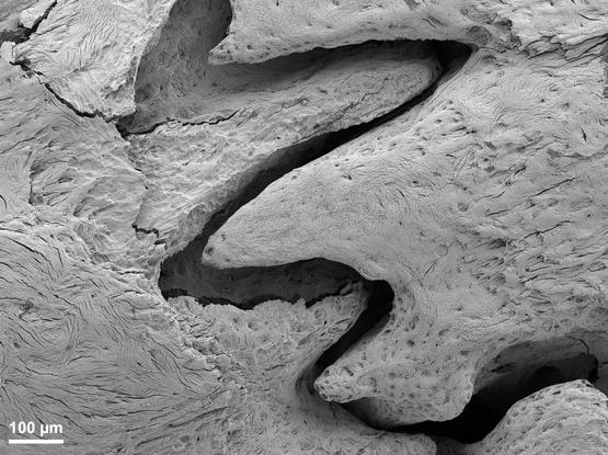
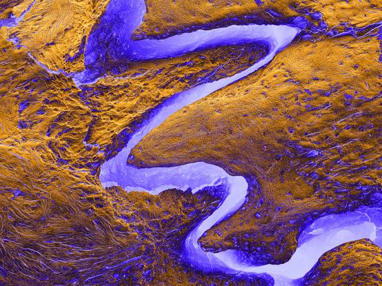
#MicroscopyMonday – No this is NOT an aerial view of the Grand Canyon.

This is a scanning electron microscopy image of the cranial sutures in the rat skull. 🔬💀🐀 These are fibrous joints where the bones of the skull interdigitate, allowing for flexibility and growth during infancy and early childhood before fusing in adulthood.

🔬🦴🧪

#bone #electronmicroscopy #imaging #biomaterials #science #medicine #biology #development #osteology #evolution

This is a scanning electron microscopy image of the cranial sutures in the rat skull. 🔬💀🐀 These are fibrous joints where the bones of the skull interdigitate, allowing for flexibility and growth during infancy and early childhood before fusing in adulthood.This is a scanning electron microscopy image of the cranial sutures in the rat skull. 🔬💀🐀 These are fibrous joints where the bones of the skull interdigitate, allowing for flexibility and growth during infancy and early childhood before fusing in adulthood.
2024-09-03

Happy #MicroscopyMonday!
Here is a kymograph of a cell crawling into a microchannel. Each horizontal row of pixels represents a line drawn from outside (green) through a channel (purple) at 5 second timepoints. ~1/2 way down (20 min) a cell fully enters the channel and takes off!

2024-08-19

Happy #MicroscopyMonday! Here are somme amoebae crawling into channels

LUT: JDM_Phase Ink Mint-Cherry

2024-07-08

🌟 Pre-vacation #MicroscopyMonday magic rendered by @Daniel_Stabley! 🌟 This is a showstopper: our top plasma membrane probes light up neurons and glia. But wait, there's more! The grand finale? A lattice light sheet stunner showing off its isotropic prowess in the XZ view. Confocal 3D rotations, eat your heart out! 🧠🔬✨ #Neuroscience #MicroscopyMagic #NextLevelImaging #cellbio #cellfie #scicomm #neuroscience #Microscopy

MEActNOWMEActNOW
2024-03-19

⚠️ That's what a leaky blood vessel looks like in the microscope. Astrocytes (🟡Gfap) around the blood vessel become reactive, inducing neuroinflammatory responses and attacking the surrounding neurons (🔵Neun) in the pathway.

2024-01-29
2023-12-11

Happy #MicroscopyMonday, from an amoeba crawling in a very narrow channel!

2023-11-06

Ich habe ein bisschen USB 3.0 Kabel zerschnitten und Querschnittsbilder mit dem Mikroskop gemacht. Das Video hat Kapitelmarken für die Eiligen und noch mehr Infos und Links in der Videobeschreibung. Viel Freude!
#Mikroskop #MicroscopyMonday #dieKleinenDinge
youtu.be/iIYT0736AQQ?feature=s

2023-10-04

Es ist zwar nicht Montag, aber ich hab lange nichts mehr zu #MicroscopyMonday gepostet, also:
Querschnitt durch ein USB3 Kabel, hier zu sehen einer der zwei Zusätzlichen DatenKanäle im USB3 Standard (lila & Orange) mit unisolierter zusätzlicher Schirmung. #Miroskop #Mikroskopie #dieKleinenDinge

Bild ist CC BY 4.0

Mikroskopieaufnahme, ca. 0,8mm x 1,6mm, Querschnitt durch ein USB3 Kabel, Detalaufnahme auf ein Kabealpaar mit lila bzw. oranger Isolation. im inneren der Isolation sind jeweils 7 Litzen aus Kupfer zu erkennen. Oberhalb verläufen zusätzliche Kupferlitzen als Abschirmung für die Datenleitung. Die Kable sind zusätzlich in einer silbrigen Folie umhüllt, an den Rändern sind noch andere Leitungen zu erahnen.
Christophe Leterrier 🔬christlet@mas.to
2023-09-18

Let's have a #notthecover for #MicroscopyMonday: hippocampal neuron with endogenously tagged HA-actin — one of the proposed covers for rupress.org/jcb/article-abstra

☆matt0815☆matt_play
2023-09-18

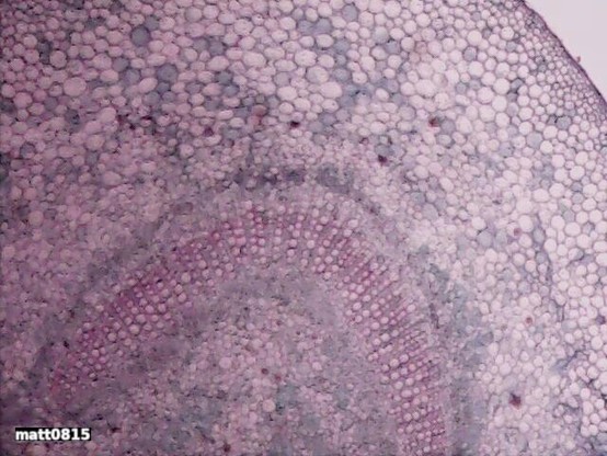
Das ist ein Blatt von einem "Nerium indicum". Dieser ist ein in Indien und Pakistan beheimateter Strauch aus der Familie der Oleander - Sehr giftig

2023-09-18

Es ist Montag, also auch #MicroscopyMonday

Im Video seht ihr eine Mikroskopie-Aufnahme von einem Mehrklingen-Rasierer, den ich langsam kippe, so dass man die Schneide-Kante einer der Klingen gut erkennen kann. Das Video stammt aus einem ganzen Mikroskopie-Stream über Messer und Klingen, hier findet ihr einen Zusammenschnitt davon: youtube.com/watch?v=9LRrt-hsuY

2023-09-11

Heute ist Montag, also auch #MicroscopyMonday

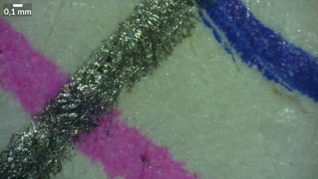
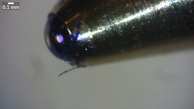
Ich habe mir mal Stifte und die entsprechenden Linien auf Papier unter dem #Mikroskop angeschaut, und das ganze sogar live gestreamt. Auf den Bildern unten seht ihr die Striche auf Papier, eine Kugelschreiber-"Spitze" und eine Nahaufnahme eines Farbdrucks. Maßstab jeweils im Bild. Bilder CC BY 4.0

Zusammenschnitt des Streams: youtu.be/sXN1yjkmCik?feature=s

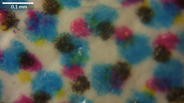
Mikroskopaufnahme 3,4mm x 1,9mm. Die Faserstruktur eines weißen Papiers ist zu erkennen, darauf rechtrs oben ein Strich mit Kugelschreiber in blau, links unten ein Strich mit einem Stabilo Point 88 in rosa und quer darüber ein Strich mit einem Bleistift.Mikroskopieaufnahme 3,4mm x 1,9mm. Nahaufnahme der Kugel an der SPitze eines Kugelschreibers. Die Kugel ist deutlich mit Tinte benetzt.Mikroskopaufnahme 0,59mm x 0,33mm. Farbdruck. Die einzelnen Farben Cyan, Magenta, Geld und Schwarz sind als gleichmäßig verteilte Punkte unterschiedlicher Größe zu erkennen
2023-09-04

Heute ist Montag, also auch #MicroscopyMonday

Ich habe mir mal die grauen Reflektorstreifen einer Warnweste unter dem Mikroskop angesehen (war in einem früheren Mikroskop-Stream auf #twitchDE) - diese Reflektorstreifen bestehen aus kleinen Glaskügelchen mit einem Durchmesser von ca. 40-70µm, eingebettet in eine Art Gel.

Bilder sind CC BY 4.0

Mikroskopieaufnahme, 1,50mm x 0,84mm es sind kleine Glaskugeln, unregelmäßig aber sehr dicht nebeneiander, teilsüberlappend angeordent, zu erkennen, Größe ca. 40-70µm.Mikroskopieaufnahme, 0,58mm x 0,32mm es sind kleine Glaskugeln zu erkennen, die durch eine graue, anscheind schmierige oder klebrige Substanz zusammen gehalten werden.

Client Info

Server: https://mastodon.social
Version: 2025.04
Repository: https://github.com/cyevgeniy/lmst