
#PhotoBioModulation #RedLightTherapy #NearInfrared #inflammation #me #cfs #logcovid #MECFS #BrainInflammation #microglia
dr. Jarred Younger on studies with near infrared light to reduce brain inflammation
#PhotoBioModulation #RedLightTherapy #NearInfrared #inflammation #me #cfs #logcovid #MECFS #BrainInflammation #microglia
dr. Jarred Younger on studies with near infrared light to reduce brain inflammation
MIT researchers invent new human brain model to enable disease research, drug discovery | MIT News
A new 3D human brain tissue platform developed by MIT researchers is the first to int…
#NewsBeep #News #Headlines #Alzheimer'sdisease #brainmodel #cerebrovasculature #dementia #extracellularmatrix(ECM) #Latvia #Li-HueiTsai #LV #microglia #MITKochInstitute #MITPicowerInstitute #MulticellularIntegratedBrains(miBrains) #neurons #personalizedmedicine #RobertLanger
https://www.newsbeep.com/231663/
Postdoc, PhD, Master
Ebner lab
Join the Ebner lab to discover lysosomal lipid logistics pathways
See the full job description on jobRxiv: https://jobrxiv.org/job/ebner-lab-27778-postdoc-phd-master/
#battendisease #lipidtransport #lysosome #membranecontactsites #microglia #microscopy #neurodegeneration #repair #ScienceJobs #hiring #research
https://jobrxiv.org/job/ebner-lab-27778-postdoc-phd-master/?fsp_sid=4848
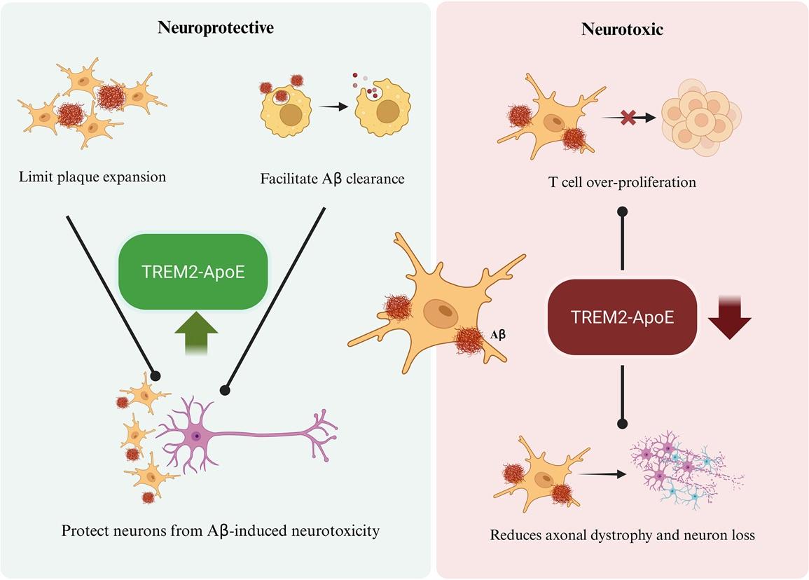
Disease-associated #microglia are a distinct state associated with #AlzheimersDisease, #ALS & #aging. This Essay explores the dynamics, origins & therapeutic potential of these microglia in #neurodegeneration, alongside evidence supporting a protective role @PLOSBiology plos.io/49mmzcm
Post-doctoral fellow
Perelman School of Medicine at the University of Pennsylvania
See the full job description on jobRxiv: https://jobrxiv.org/job/perelman-school-of-medicine-at-the-university-of-pennsylvania-27778-post-doctoral-fellow/
#3Dcellculture #animalmodels #braindiseases #macrophage #microglia #ScienceJobs #hiring #research
https://jobrxiv.org/job/perelman-school-of-medicine-at-the-university-of-pennsylvania-27778-post-doctoral-fellow/?fsp_sid=4190
Postdoc, PhD, Master
Ebner lab
Join the Ebner lab to discover lysosomal lipid logistics pathways
See the full job description on jobRxiv: https://jobrxiv.org/job/ebner-lab-27778-postdoc-phd-master/
#battendisease #lipidtransport #lysosome #membranecontactsites #microglia #microscopy #neurodegeneration #repair #ScienceJobs #hiring #research
https://jobrxiv.org/job/ebner-lab-27778-postdoc-phd-master/?fsp_sid=3455
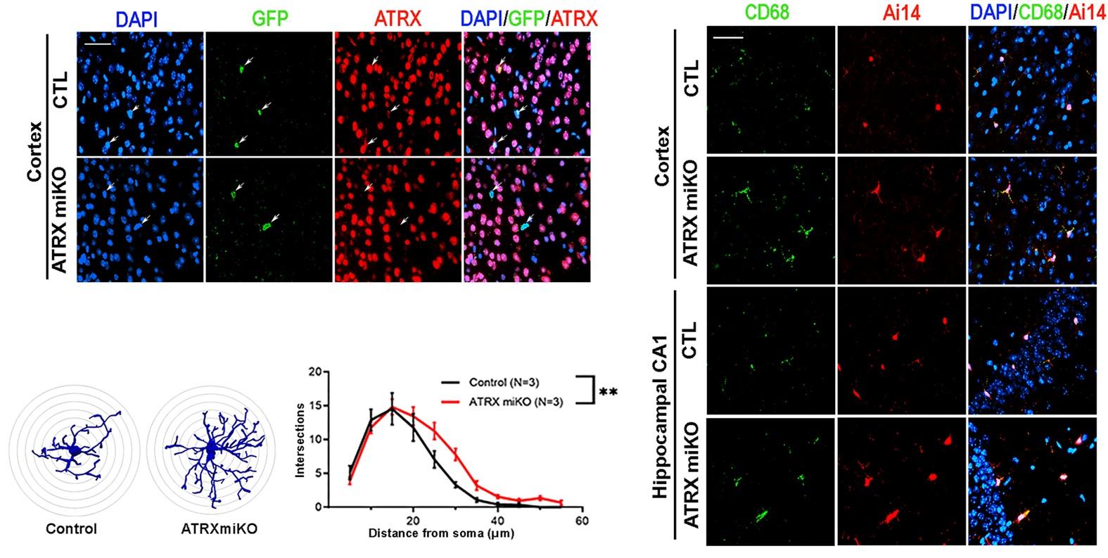
What's the role of microglial chromatin-mediated processes? This study shows that loss of ATRX in #microglia disrupts #chromatin structure, leading to de-repression of #retroelements & a viral mimicry #inflammation response that impairs hippocampal neuron function @PLOSBiology https://plos.io/46cBaE4
Post-doctoral fellow
Perelman School of Medicine at the University of Pennsylvania
See the full job description on jobRxiv: https://jobrxiv.org/job/perelman-school-of-medicine-at-the-university-of-pennsylvania-27778-post-doctoral-fellow/
#3Dcellculture #animalmodels #braindiseases #macrophage #microglia #ScienceJobs #hiring #research
https://jobrxiv.org/job/perelman-school-of-medicine-at-the-university-of-pennsylvania-27778-post-doctoral-fellow/?fsp_sid=2265
Circadian Harmony: Aligning Sleep-Wake Cycles for Brain Health
#SleepScience #BrainHealth #CircadianRhythm #SleepStages #Memory #Neuroinflammation #Glia #Microglia #Neurodegeneration #Melatonin #Serotonin #SleepBetter
Postdoc, PhD, Master
Ebner lab
Join the Ebner lab to discover lysosomal lipid logistics pathways
See the full job description on jobRxiv: https://jobrxiv.org/job/ebner-lab-27778-postdoc-phd-master/
#battendisease #lipidtransport #lysosome #membranecontactsites #microglia #microscopy #neurodegeneration #repair #ScienceJobs #hiring #research
https://jobrxiv.org/job/ebner-lab-27778-postdoc-phd-master/?fsp_sid=1096
How Your Immune System and Brain Communicate: The Hidden Connection
#ImmuneSystem #BrainHealth #Microglia #Inflammation #MentalHealth #Cytokines #StressAndMood #Neuroscience #InflammationAndDepression #Neuroimmunology
Understanding Brain Trauma: Cellular Responses and Molecular Cascades
#BrainInjury #Neuroinflammation #TraumaticBrainInjury #Microglia #Astrocytes #BloodBrainBarrier #Neuroregeneration #BrainHealth #Neuroscience #BrainRecovery
From Trauma to Treatment: Navigating Brain Injury and Inflammation
#BrainInjury #Neuroinflammation #Concussion #BrainHealth #Microglia #NeuroScience #BrainRecovery #Neurodegeneration #MentalHealth #NeuroscienceResearch
When Brain Balance Tips: Understanding Neuroinflammation After Head Injuries
#BrainHealth #Microglia #Neuroinflammation #Neurodegeneration #Alzheimers #Parkinsons #BrainScience #Neuroscience #BrainAging #Neurobiology #ProteinAggregates #BrainRepair #Neuroimmunology #BrainCells #CognitiveHealth
Tryptophan and the Kynurenine Pathway: Metabolic Routes Impacting Mood and Neurodegeneration
#BrainChemistry #Neuroimmune #KynureninePathway #GutBrainAxis #Neuroinflammation #Microglia #Astrocytes #Neurodegeneration #OxidativeStress #MentalHealth #BrainHealth #Metabolism #NervousSystem #DietAndBrain #Neuroscience
Postdoc, PhD, Master
Ebner lab
Join the Ebner lab to discover lysosomal lipid logistics pathways
See the full job description on jobRxiv: https://jobrxiv.org/job/ebner-lab-27778-postdoc-phd-master/
#battendisease #lipidtransport #lysosome #membranecontactsites #microglia #microscopy #neurodegeneration #repair #ScienceJobs #hiring #research
https://jobrxiv.org/job/ebner-lab-27778-postdoc-phd-master/?fsp_sid=39
Cellular Heroes of the Brain: Microglia, Astrocytes, and More
#Neuroscience #BrainHealth #Neuroplasticity #Neurobiology #BrainScience #Neurodegeneration #Microglia #Astrocytes #Memory #BrainTips
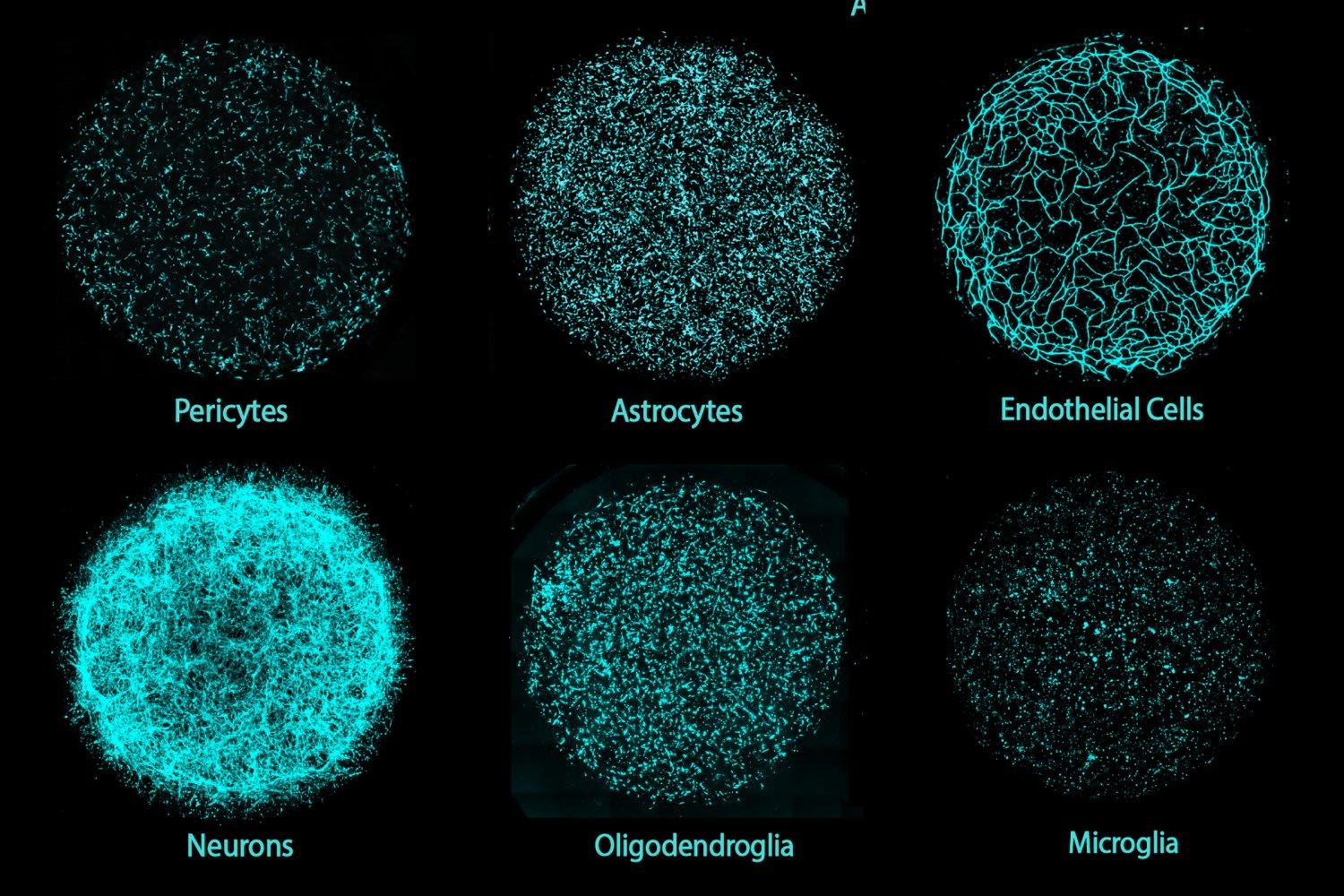
Neurons, Glia, and Blood Vessels: The Complex City of the Brain
#BrainHealth #Neuroscience #AlzheimersDisease #Neuroinflammation #Microglia #CognitiveHealth #Neurodegeneration #BrainResearch #NeurotrophicFactors #HealthyBrain #MindAndMemory #BrainScience #NeuralNetworks #BrainCare #Neuroprotection
The Brain’s Tiny Guardians: Microglia and Neuroinflammation Explained
#Microglia #Neurodegeneration #BrainHealth #Alzheimers #Parkinsons #Neuroinflammation #CognitiveHealth #BrainScience #Neuroscience #Neurotherapy
The Brain's Metropolis: How Microglia, Gut Microbes, and Stress Shape Your Mind
#BrainHealth #Neuroinflammation #GutBrainAxis #Microglia #MentalHealth #Neuroplasticity #GutMicrobiome #StressAndBrain #NeuroScience #BrainTour